

Oxidative damage in brain from human mutant APP/PS-1 double knock-in mice as a function of age
- PMID: 18762245
- PMCID: PMC2597707
- DOI: 10.1016/j.freeradbiomed.2008.08.012
Oxidative damage in brain from human mutant APP/PS-1 double knock-in mice as a function of age
Abstract
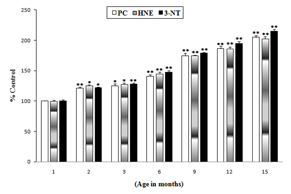
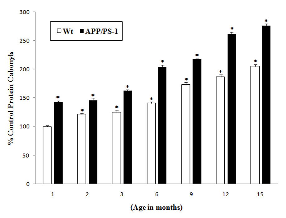
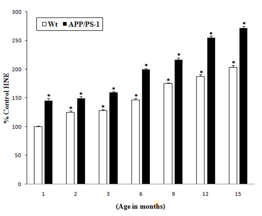
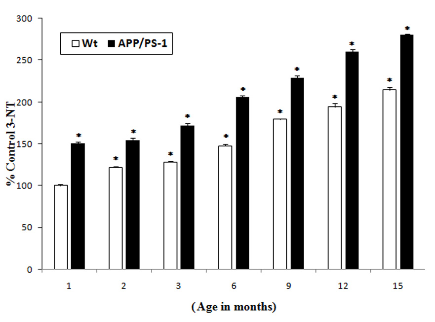
Oxidative stress is strongly implicated in the progressive decline of cognition associated with aging and neurodegenerative disorders. In the brain, free radical-mediated oxidative stress plays a critical role in the age-related decline of cellular function as a result of the oxidation of proteins, lipids, and nucleic acids. A number of studies indicate that an increase in protein oxidation and lipid peroxidation is associated with age-related neurodegenerative diseases and cellular dysfunction observed in aging brains. Oxidative stress is one of the important factors contributing to Alzheimer's disease (AD), one of whose major hallmarks includes brain depositions of amyloid beta-peptide (Abeta) derived from amyloid precursor protein (APP). Mutation in APP and PS-1 genes, which increases production of the highly amyloidogenic amyloid beta-peptide (Abeta42), is the major cause of familial AD. In the present study, protein oxidation and lipid peroxidation in the brain from knock-in mice expressing human mutant APP and PS-1 were compared with brain from wild type, as a function of age. The results suggest that there is an increased oxidative stress in the brain of wild-type mice as a function of age. In APP/PS-1 mouse brain, there is a basal increase (at 1 month) in oxidative stress compared to the wild type (1 month), as measured by protein oxidation and lipid peroxidation. In addition, age-related elevation of oxidative damage was observed in APP/PS-1 mice brain compared to that of wild-type mice brain. These results are discussed with reference to the importance of Abeta42-associated oxidative stress in the pathogenesis of AD.
Figures
References
-
- Selkoe DJ. Aging, amyloid, and Alzheimer's disease: a perspective in honor of Carl Cotman. Neurochem Res. 2003;28:1705–1713. - PubMed
-
- Butterfield DA, Drake J, Pocernich C, Castegna A. Evidence of oxidative damage in Alzheimer's disease brain: central role for amyloid beta-peptide. Trends Mol Med. 2001;7:548–554. - PubMed
-
- Butterfield DA, Lauderback CM. Lipid peroxidation and protein oxidation in Alzheimer's disease brain: potential causes and consequences involving amyloid beta-peptide-associated free radical oxidative stress. Free Radic Biol Med. 2002;32:1050–1060. - PubMed
-
- Markesbery WR. Oxidative stress hypothesis in Alzheimer's disease. Free Radic Biol Med. 1997;23:134–147. - PubMed
-
- Mattson MP. Oxidative stress, perturbed calcium homeostasis, and immune dysfunction in Alzheimer's disease. J Neurovirol. 2002;8:539–550. - PubMed
Publication types
MeSH terms
Substances
Grants and funding
LinkOut - more resources
Full Text Sources
Other Literature Sources
Medical
Molecular Biology Databases
