. 2008 Sep 29;205(10):2269-80.
doi: 10.1084/jem.20071371. Epub 2008 Sep 1.

Delayed maturation of an IL-12-producing dendritic cell subset explains the early Th2 bias in neonatal immunity

Affiliations

Delayed maturation of an IL-12-producing dendritic cell subset explains the early Th2 bias in neonatal immunity

Hyun-Hee Lee et al. J Exp Med. .

Abstract

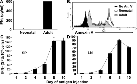
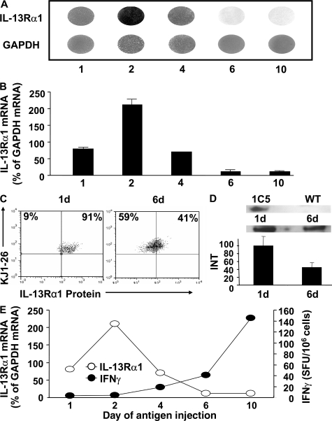
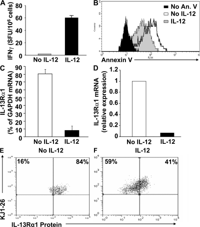
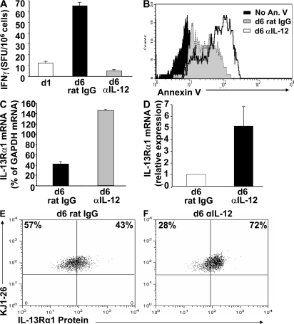
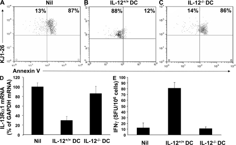
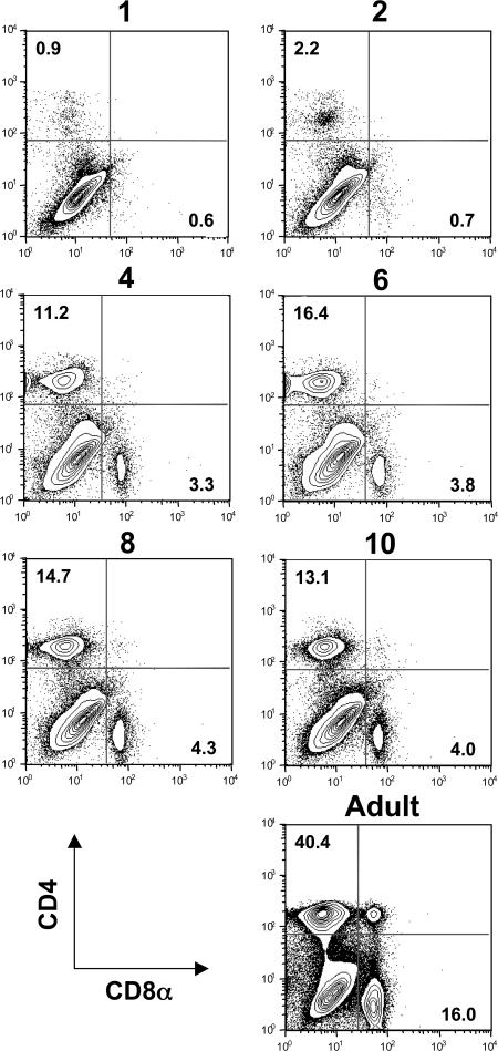
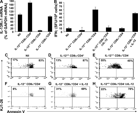
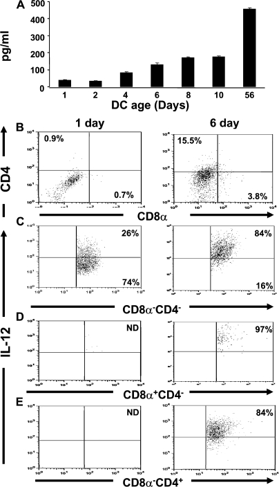
Primary neonatal T cell responses comprise both T helper (Th) cell subsets, but Th1 cells express high levels of interleukin 13 receptor alpha1 (IL-13R alpha 1), which heterodimerizes with IL-4R alpha. During secondary antigen challenge, Th2-produced IL-4 triggers the apoptosis of Th1 cells via IL-4R alpha/IL-13R alpha 1, thus explaining the Th2 bias in neonates. We show that neonates acquire the ability to overcome the Th2 bias and generate Th1 responses starting 6 d after birth. This transition was caused by the developmental maturation of CD8 alpha(+)CD4(-) dendritic cells (DCs), which were minimal in number during the first few days of birth and produced low levels of IL-12. This lack of IL-12 sustained the expression of IL-13R alpha 1 on Th1 cells. By day 6 after birth, however, a significant number of CD8 alpha(+)CD4(-) DCs accumulated in the spleen and produced IL-12, which triggered the down-regulation of IL-13R alpha 1 expression on Th1 cells, thus protecting them against IL-4-driven apoptosis.


Figures

Figure 1.
Figure 1.
Neonates acquire the ability to develop secondary Th1 responses when the primary encounter with Ag occurs on or beyond day 6 after birth. Newborn BALB/c mice were given 3 × 10⁴ neonatal or adult DO11.10 CD4 T cells and injected with 100 μg Ig-OVA in saline, and 2 wk later, the splenic cells (106 cells per well) were stimulated with 10 μM OVA peptide in vitro. (A) Production of IFN-γ was measured by ELISA. (B) Apoptosis of IFN-γ–producing KJ1-26+ Th1 cells was evaluated by Annexin V staining. In B, 10 μg/ml BFA was added after 6 h of stimulation with OVA peptide, and the culture was continued for another 6 h to facilitate intracellular accumulation of IFN-γ. The cells were then labeled with KJ1-26 and Annexin V, and stained for intracellular IFN-γ. Histogram shows gating on KJ1-26+/ IFN-γ+ cells. (C and D) Newborn BALB/c mice were given 3 × 10⁴ neonatal DO11.10 T cells within 24 h after birth and were injected with 60 μg/g Ig-OVA (to adjust for growth) on day 1, 2, 4, 6, 8, or 10 after T cell transfer. 2 mo later, the mice were challenged with 125 μg OVA peptide in CFA. 10 d later, the splenic (106 cells/well) and lymph node (0.5 × 10⁶ cells/well) cells were stimulated with 10 μM OVA peptide for 24 h, and production of IFN-γ was determined by ELISPOT. Each bar represents the mean ± SD of triplicate wells. The data are representative of two experiments.
Figure 2.
Figure 2.
Recovery of neonatal IFN-γ response coincides with down-regulation of IL-13Rα1 expression. Newborn BALB/c mice were given 3 × 10⁵ purified neonatal CD4+ DO11.10 T cells and injected i.p. with 60 μg/g Ig-OVA in saline at day 1, 2, 4, 6, or 10 after birth, and 2 wk later, the splenic cells (107 cells/ml) were stimulated for 10 h with 10 μM OVA peptide. (A) Subsequently, Th1 cells were isolated, and RNA was extracted and used for analysis of IL-13Rα1 gene expression as well as GAPDH control by spot-blot technology, as described in Materials and methods. (B) The intensity of radioactive spots was analyzed on a Molecular Imager FX using Quantity One software and presented as a ratio of IL-13Rα1 to GAPDH after deduction of the background intensity obtained with pUC19 DNA negative control. The bars represent the mean ± SD of two experiments. (C and D) Newborn BALB/c mice were given 3 × 10⁴ neonatal CD4+ DO11.10 T cells and injected i.p. with 60 μg/g Ig-OVA in saline at day 1 or 6 after birth, and 2 wk later, the splenic cells (106 cells/ml) were stimulated for 10 h with 10 μM OVA peptide. In C, the cells were stained with KJ1-26 and rabbit anti–IL-13Rα1 antiserum (1:100 dilution), and Th1 cells were identified by staining for intracellular IFN-γ with anti–IFN-γ antibody. Expression of surface IL-13Rα1 was determined by flow cytometry on cells gated for KJ1-26 expression and intracellular IFN-γ production. In D, Th1 cells were purified as in A, lysed using NP-40 detergent, and run on a 10% acrylamide gel. Transfer was made onto a nitrocellulose membrane, and IL-13Rα1 protein was detected using rabbit anti–IL-13Rα1 antibodies (1:1,000 dilution). For control purposes, we used lysates from CTLL-2 cells transfected with a plasmid coding for cell-surface IL-13Rα1 (reference 17) or wild-type untransfected CTLL-2 cells. Band intensity was analyzed using the Molecular Imager FX, and the relative expression of IL-13Rα1 represents a percent ratio with 1 d as 100%. Each bar represents the mean ± SD of duplicate samples. (E) IL-13Rα1 mRNA expression measured by spot intensity from the experiments in A and B is illustrated together with the IFN-γ production presented in Fig. 1 C to correlate IL-13Rα1 down-regulation together with an increase in secondary IFN-γ production.
Figure 3.
Figure 3.
Exogenous IL-12 inhibits developmental IL-13Rα1 up-regulation and restores secondary Th1 IFN-γ response in vivo. Newborn BALB/c mice were given 3 × 10⁴ neonatal DO11.10 T cells within 24 h after birth. 1 d later, the recipient mice were given 100 μg Ig-OVA and 50 ng rIL-12. The mice were given additional IL-12 on days 2 and 3 after T cell transfer. 7 wk later, the mice were challenged with 125 μg OVA peptide in CFA, and 10 d after the challenge, the splenic cells (106 cells/well) were stimulated for 24 h with 10 μM OVA peptide. (A and B) Subsequently, IFN-γ was measured by ELISPOT (A) and apoptosis was evaluated using Annexin V staining of KJ1-26+/IFN-γ+ splenic cells (B). For Annexin V staining, 10 μg/ml BFA was added during the last 8 h of peptide stimulation. Each bar in A represents the mean ± SD of triplicate wells. (C and D) Mice that received neonatal DO11.10 T cells were given Ig-OVA and IL-12 as in A, and 2 wk later, Th1 cells were isolated and IL-13Rα1 expression was assessed by spot blot (C) and real-time PCR (D). The real-time PCR used 200 ng RNA and the Absolute QRT-PCR SYBR kit to determine IL-13Rα1 mRNA. For the spot blot in C, each bar represents the mean ± SD of duplicate spots. For the real-time PCR, the bars represent the comparative threshold cycle (CT). The value of the sample from mice not receiving IL-12 was set as 1. (E and F) Newborn BALB/c mice given 3 × 10⁴ neonatal CD4+ DO11.10 T cells were exposed to Ig-OVA in the presence of IL-12 as in A and B, and 2 wk later, the splenic cells (106 cells/ml) were stimulated for 10 h with 10 μM OVA peptide. The cells were then stained with KJ1-26 and rabbit anti–IL-13Rα1 antiserum (1:100 dilution), and Th1 cells were identified by staining for intracellular IFN-γ with anti–IFN-γ antibody. Expression of surface IL-13Rα1 was determined by flow cytometry on cells gated for KJ1-26 expression and intracellular IFN-γ production.
Figure 4.
Figure 4.
Neutralization of IL-12 on day 6 after birth nullifies IFN-γ response through up-regulation of IL-13Rα1 and apoptosis of Th1 cells. Newborn BALB/c mice were given 3 x 10⁴ CD4 neonatal DO11.10 T cells within 24 h after birth. On day 6, the hosts were given 60 μg/g Ig-OVA and 50 μg anti–IL-12 antibody per mouse or rat IgG. Another injection of anti–IL-12 antibody or rat IgG was given on days 7 and 8. A group of mice that received Ig-OVA on day 1 and no anti–IL-12 antibody at any time was included as a control. 7 wk later, all groups were challenged with 125 μg OVA peptide in CFA. 10 d later, the splenic cells (106 cells/well) were stimulated with 10 μM OVA peptide and assayed for (A) IFN-γ production by ELISPOT and (B) apoptosis by Annexin V staining, as in Fig. 3. Each bar represents the mean ± SD of triplicate wells. (C and D) Mice that received neonatal DO11.10 T cells were given Ig-OVA and anti–IL-12 antibody as in A, and 2 wk later, primary Th1 cells were isolated and IL-13Rα1 expression was assessed by spot blot (C) and real-time PCR (D), as described in Fig. 3. For the spot blot in C, each bar represents the mean ± SD of duplicate spots. For the real-time PCR, the bars represent the comparative threshold cycle (CT). The value of the sample from mice that received rat IgG was set as 1. (E and F) Newborn BALB/c from A and B that received anti–IL-12 antibody or rat IgG control during exposure to Ig-OVA on day 6 were killed 2 wk later, and the splenic cells were stained with KJ1-26 and rabbit anti–IL-13Rα1 antiserum (1:100 dilution). The Th1 cells were identified by staining for intracellular IFN-γ with anti–IFN-γ antibody, and expression of surface IL-13Rα1 was determined by flow cytometry on cells gated for KJ1-26 expression and intracellular IFN-γ production.
Figure 5.
Figure 5.
Transfer of adult IL-12+/+ but not IL-12−/− DCs into newborn mice down-regulates IL-13Rα1 expression, inhibits apoptosis, and restores secondary Th1 IFN-γ responses. Newborn (1-d-old) BALB/c mice were given 3 × 10⁴ neonatal DO11.10 T cells without (Nil) or with 2 × 10⁵ splenic DCs from adult wild-type (IL-12+/+ DCs) or IL-12–deficient (IL-12−/− DCs) BALB/c mice (reference 20) and were injected 1 d later with 100 μg Ig-OVA. After 7 wk, the mice were challenged with 125 μg OVA peptide in CFA, and 10 d later, the splenic cells (106 cells/well) were stimulated with 10 μM OVA peptide for 24 h. (A–C) Apoptosis of KJ1-26+/IFN-γ+ Th1 cells was evaluated by staining with Annexin V, as described in Fig. 3. (D) Groups of mice were killed 2 wk after T cell/DC transfer and exposure to Ig-OVA, and primary Th1 cells were isolated using IFN-γ secretion kit, as described in Materials and methods. RNA was then extracted and IL-13Rα1 expression was determined by spot blot, as in Fig. 2. Because IL-13Rα1 mRNA parallels with protein expression (see Figs. 2, 3, and 4), all follow-up experiments will use mRNA analysis on separated Th1 cells. (E) IFN-γ production was measured using ELISPOT assay. Each bar represents the mean ± SD of duplicate spots.
Figure 6.
Figure 6.
Developmental accumulation of CD8α+CD4 and CD8αCD4+ DCs occurs at day 6 after birth. Splenic cells (106 cells/ tube) from 1-, 2-, 4-, 6-, 8-, and 10-d-old BALB/c neonates were stained with anti-CD11c, anti-CD4, and anti-CD8α antibodies, fixed with 2% formaldehyde, and analyzed by FACS. Expression of CD4 and CD8α was analyzed on cells gated on CD11c. Splenic cells from adult (8-wk-old) BALB/c mice were included as control. Percentages of cells are shown.
Figure 7.
Figure 7.
The CD8α+CD4 but not the CD8αCD4+ DC subset inhibits up-regulation of IL-13Rα1, prevents apoptosis, and restores neonatal IFN-γ responses. Newborn BALB/c mice were given neonatal DO11.10 T cells without (Nil) or with 105 CD8α+CD4 or CD8αCD4+ DCs from adult IL-12+/+ or IL-12−/− BALB/c mice and were injected 24 h later with 100 μg Ig-OVA. (A) The mice were killed within 2 wk, primary Th1 cells were isolated on the basis of IFN-γ secretion, and IL-13Rα1 expression was evaluated by spot blot. Each bar represents the mean ± SD of duplicate spots. (B–H) 7 wk after exposure to Ig-OVA the mice were challenged with 125 μg OVA peptide in CFA. 10 d later, the splenic cells (106 cells/well) were stimulated for 24 h with 10 μM OVA peptide, and production of IFN-γ was measured by ELISPOT (B) and apoptosis was evaluated by Annexin V staining of KJ1-26+/IFN-γ+ splenic cells (C–H). For neutralization of endogenous IL-12 where indicated (IL-12+/+ CD8α+CD4/anti–IL-12), the mice were given i.p. 50 μg anti–IL-12 antibody (clone C17.8) on days 1, 2, and 3 after neonatal transfer of DO11.10 T cells and DCs. For stimulation with OVA peptide in the presence of rIL-12 where indicated (IL-12−/−CD8α+CD4 + IL-12), the culture was supplemented with 2 ng/ml rIL-12. Each bar in B represents the mean ± SD of triplicate wells.
Figure 8.
Figure 8.
The limited availability of IL-12 during neonatal exposure to Ag is related to delayed accumulation in the spleen of the presenting CD8α+CD4 DC subset. Newborn BALB/c mice were killed at the indicated day after birth, and splenic CD11c+ DCs were isolated on anti-CD11c microbeads. (A) The DCs (100 × 103 cells per well) were stimulated with 6 μg/ml CpG, and 24 h later, IL-12 was measured by ELISA. Each bar represents the mean ± SD of triplicate wells. (B–E) 1- and 6-d-old DCs (100 × 103 cells per well) were purified from the spleens of 45 newborns, and 106 CD11c+ cells were stimulated with 6 μg/ml CpG for 10 h, after which BFA was added and the culture was continued for 4 h to facilitate intracellular cytokine accumulation. Subsequently, the cells were stained with anti-CD11c, anti-CD4, and anti-CD8 antibodies and permeabilized with 2% saponin. Intracellular IL-12 was detected by staining with anti–IL-12 p70 antibody. B shows the frequency of the subsets, and C–E illustrates flow cytometry plots of intracellular IL-12 on the indicated subset. ND, not detected.

References

    1. Singh, R.R., B.H. Hahn, and E.E. Sercarz. 1996. Neonatal peptide exposure can prime T cells and, upon subsequent immunization, induce their immune deviation: implications for antibody vs. T cell-mediated autoimmunity. J. Exp. Med. 183:1613–1621. - PMC - PubMed
    1. Chen, N., and E.H. Field. 1995. Enhanced type 2 and diminished type 1 cytokines in neonatal tolerance. Transplantation. 59:933–941. - PubMed
    1. Forsthuber, T., H.C. Yip, and P.V. Lehmann. 1996. Induction of TH1 and TH2 immunity in neonatal mice. Science. 271:1728–1730. - PubMed
    1. Min, B., K.L. Legge, C. Pack, and H. Zaghouani. 1998. Neonatal exposure to a self-peptide–immunoglobulin chimera circumvents the use of adjuvant and confers resistance to autoimmune disease by a novel mechanism involving interleukin 4 lymph node deviation and interferon γ–mediated splenic anergy. J. Exp. Med. 188:2007–2017. - PMC - PubMed
    1. Adkins, B., C. Leclerc, and S. Marshall-Clarke. 2004. Neonatal adaptive immunity comes of age. Nat. Rev. Immunol. 4:553–564. - PubMed

Publication types

MeSH terms