. 2008 Sep 8;182(5):979-91.
doi: 10.1083/jcb.200712110. Epub 2008 Sep 1.

IGFBP-5 regulates muscle cell differentiation by binding to IGF-II and switching on the IGF-II auto-regulation loop

Affiliations

IGFBP-5 regulates muscle cell differentiation by binding to IGF-II and switching on the IGF-II auto-regulation loop

Hongxia Ren et al. J Cell Biol. .

Abstract

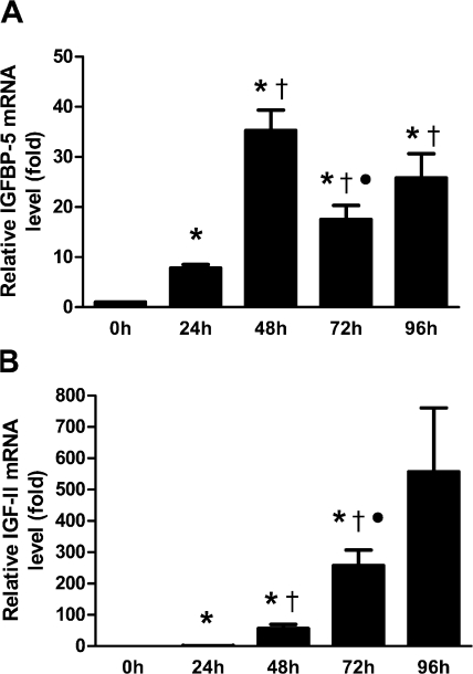
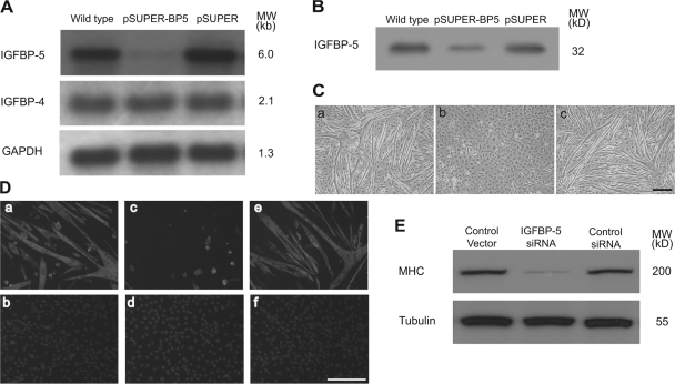
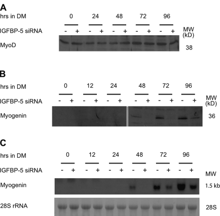
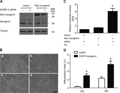
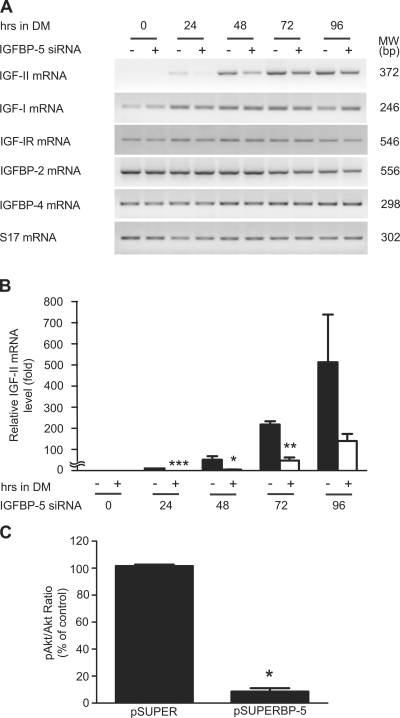
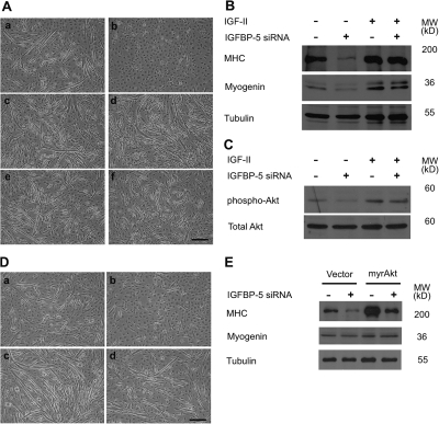
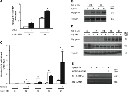
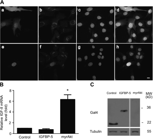
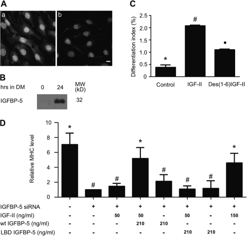
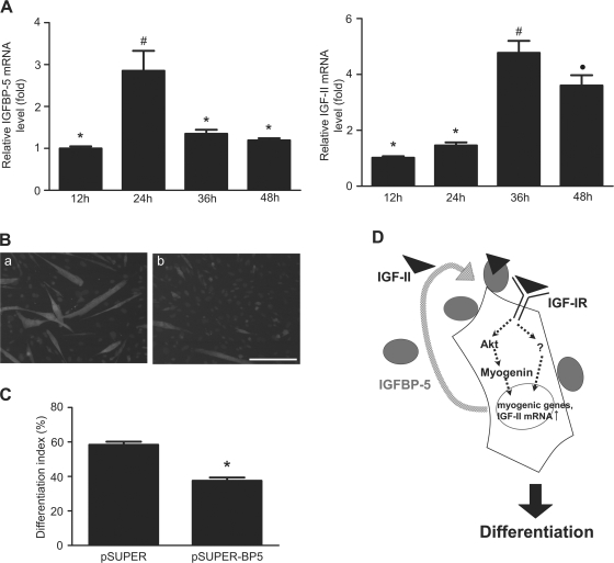
IGF-II stimulates both mitogenesis and myogenesis through its binding and activation of the IGF-I receptor (IGF-IR). How this growth factor pathway promotes these two opposite cellular responses is not well understood. We investigate whether local IGF binding protein-5 (IGFBP-5) promotes the myogenic action of IGF-II. IGFBP-5 is induced before the elevation of IGF-II expression during myogenesis. Knockdown of IGFBP-5 impairs myogenesis and suppresses IGF-II gene expression. IGF-II up-regulates its own gene expression via the PI3K-Akt signaling pathway. Adding IGF-II or constitutively activating Akt rescues the IGFBP-5 knockdown-caused defects. However, an IGF analogue that binds to the IGF-IR but not IGFBP has only a limited effect. When added with low concentrations of IGF-II, IGFBP-5 restores IGF-II expression and myogenic differentiation, whereas an IGF binding-deficient IGFBP-5 mutant has no effect. These findings suggest that IGFBP-5 promotes muscle cell differentiation by binding to and switching on the IGF-II auto-regulation loop.


Figures

Figure 1.
Figure 1.
The induction of IGFBP-5 precedes that of IGF-II during myogenesis. Cultured C2C12 myoblasts were induced to differentiate by switching to differentiation medium (DM). Total RNA was extracted at different time points. IGFBP-5 mRNA (A) and IGF-II mRNA (B) levels were measured by qRT-PCR. Values are expressed as relative levels to that of 24 h for IGF-II (IGF-II mRNA levels were under the detection limit at 0 h) or to that of the 0 h group for IGFBP-5 after normalized to cyclophilin levels. Data shown are means ± SE of three independent experiments. *, P < 0.05, compared with the 0 h group; †, P < 0.05, compared with the 24-h group; •, P < 0.05, compared with the 48 h group.
Figure 2.
Figure 2.
Knockdown of IGFBP-5 impairs myogenic differentiation. (A) C2C12 cells were transfected with pSUPER or pSUPER-BP5. 3 d later, total RNA was isolated and subjected to Northern blot analysis. (B) Conditioned media obtained from wild-type, pSUPER-BP5–transfected, and pSUPER-transfected cells were subjected to ligand blot analysis. (C) Knockdown of IGFBP-5 inhibits myotube formation. C2C12 cells were transfected with the pSUPER vector (a), pSUPER-BP5 (b), or control pSUPER (c). 30 h after transfection, cells were induced to differentiate for 4 d. Images were representative from three reproducible experiments. Bar, 200 μm. (D) Knockdown of IGFBP-5 reduces MHC expression. Cells described in C were analyzed by immunostaining for MHC (a, c, and e) and counterstained with DAPI (b, d, and f). Bar, 200 μm. (E) Western immunoblot analysis of cells described in C.
Figure 3.
Figure 3.
Knockdown of IGFBP-5 decreases Myogenin expression. After transfection, C2C12 myoblasts were switched to DM. Samples were collected at the indicated time. (A) Western immunoblot of MyoD. (B) Western immunoblot analysis of Myogenin. (C) Northern blot analysis of myogenin.
Figure 4.
Figure 4.
Forced expression of Myogenin does not rescue the myogenic defects in IGFBP-5–deficient cells. (A) Western immunoblot analysis of C2C12 cells transfected with pSUPER + pCS2, pSUPER-BP5 + pCS2, pSUPER + Myc-tagged Myogenin, or pSUPER-BP5 + Myc-tagged Myogenin 4 d after they were induced to differentiate. (B) Phase-contrast images of cells transfected with pSUPER + pCS2 (a), pSUPER-BP5 + pCS2 (b), pSUPER + Myc-tagged Myogenin (c), and pSUPER-BP5 + Myc-tagged (d). These were representative images from four reproducible experiments. Bar, 200 μm. (C) Luciferase activity was measured 2 d after differentiation was induced in C2C12 cells transfected with an empty vector and the Myogenin reporter construct 4RTK, Myc-tagged Myogenin and the reporter construct TK, and overexpression of Myogenin and 4RTK, respectively. Data are expressed as relative value to those of the empty vector and 4RTK transfected cells. Data shown are means ± SE of three independent experiments. *, P < 0.05. (D) C2C12 cells were transfected with either EGFP or EGFP-tagged Myogenin and induced to differentiate for 24 or 36 h, and their differentiation indexes were determined. Data shown are means ± SE of two independent experiments each performed in triplicate. *, P < 0.05.
Figure 5.
Figure 5.
Knockdown of IGFBP-5 suppresses IGF-II gene expression and decreases IGF-IR–mediated signaling activity. (A) C2C12 cells transfected with pSUPER or pSUPER-BP5 were induced to differentiate by switching to DM. RNA samples were prepared at the indicated time points and subjected to RT-PCR. (B) RNA samples described in A were analyzed by qRT-PCR. Data are expressed as relative value to those of the pSUPER transfected cells at 24 h after the induction of differentiation. Data shown are means ± SE of three independent experiments. *, P < 0.05; **, P < 0.01; ***, P < 0.001 compared with the pSUPER-transfected control groups at the same time point. (C) Cells transfected with pSUPER or pSUPER-BP5 was induced to differentiate. 48 h later, cells were lysed and analyzed by Western immunoblot. The phospho-Akt/total Akt ratio was calculated by densitometry. Values are expressed as relative levels to pSUPER group. Data shown are means ± SE; n = 4; *, P < 0.05.
Figure 6.
Figure 6.
Exogenous IGFs or expression of a constitutively active Akt “rescues” the myogenic defects caused by IGFBP-5 knockdown. (A) Effects of IGFs. C2C12 cells transfected with pSUPER (a, c, and e) or pSUPER-BP5 (b, d, and f) were induced to differentiate by switching to the DM containing 0.5% horse serum without IGF-I (a and b), or with 400 ng/ml IGF-I (c and d), or 400 ng/ml IGF-II (e and f) for 4 d. Representative images from four reproducible experiments are shown. Bar, 200 μm. (B) Western immunoblot analysis of cell lysates from the groups indicated. (C) Western immunoblot analysis of Akt in cell lysates from the groups indicated. (D) Effect of myrAkt. C2C12 cells transfected with pSUPER + pCS2 (a), pSUPER-BP5 + pCS2 (b), pSUPER + pCS2+myr-Akt (c), and pSUPER-BP5 + pCS2+myr-Akt (d) were switched to the DM containing 0.5% horse serum for 4 d. Phase-contrast images were representative images from two independent experiments. Bar, 200 μm. (E) Western immunoblot analysis of cell lysates from the groups indicated.
Figure 7.
Figure 7.
IGF-II up-regulates its own gene expression through the PI3K-Akt signaling pathway. (A) Wild-type C2C12 cells were switched to SFM supplemented with or without 300 ng/ml IGF-II. IGF-II mRNA levels were measured by qRT-PCR and normalized by cyclophilin mRNA levels. Data shown represent means ± SE of two independent experiments. *, P < 0.05 compared with the control group at the same time point. (B) Wild-type C2C12 cells were switched to DM (containing 0.5% horse serum) supplemented with or without 300 ng/ml IGF-II. 24 and 36 h later, cells were lysed and subjected to Western blot analysis. (C) Cells transfected with the empty pCS2 (open box) or pCS2+myr-Akt plasmid (closed box) were switched to DM. IGF-II mRNA levels were measured by qRT-PCR and normalized. The results were expressed as relative value to those of the empty vector transfected cells at 0 h. Data shown are means ± SE of three independent experiments. *, P < 0.05; **, P < 0.01 compared with the control group at the same time point. (D) C2C12 cells transfected with the empty pCS2+ or pCS2+myr-Akt plasmid were switched to the DM. Cells were lysed at the indicated time and subjected to Western blot analysis. (E) Overexpression of Myogenin does not restore the reduced IGF-II expression in IGFBP-5 knocked down cells. Cells transfected with pSUPER + pCS2, pSUPER + Myc-Myogenin, pSUPER-BP5 + pCS2, and pSUPER-BP5 + Myc-Myogenin were induced to differentiate for 4 d. RNA samples were prepared and subjected to RT-PCR analysis.
Figure 8.
Figure 8.
IGFBP-5 does not exert direct effect on IGF-II gene expression. (A) IGFBP-5 is not localized in the nucleus. C2C12 cells were transfected with either pcDNA3.1-EGFP (a–d) or pcDNA3.1-EGFP-mIGFBP-5 (e–h). 24 h after the induction of differentiation, cells were fixed, the membrane permeabilized, and stained with an IGFBP-5 antibody (b and f) and counter-stained with DAPI (c and g). Panels a and e are GFP signal, and panels d and h are overlays. Bar, 10 μm. (B) Forced nuclear expression of the IGFBP-5 transactivation domain does not increase IGF-II expression. C2C12 cells were transfected with either pBIND vector, pBIND-BP5, or myrAkt and induced to differentiate. 36 h later, total RNA samples were isolated and IGF-II mRNA levels were determined by qRT-PCR. Values are relative to the control group after normalized by cyclophilin levels. *, P < 0.05 compared with the control group. (C) Western blot of cell lysates described in B using a Gal4 antibody.
Figure 9.
Figure 9.
IGFBP-5 is localized on the cell surface and promotes myogenic differentiation by binding to and promoting IGF-II action. (A) 24 h after the induction of differentiation, C2C12 cells were fixed without (a) or with (b) membrane permeabilization and stained with an IGFBP-5 antibody. Bar, 10 μm. (B) 0 and 24 h after induction of differentiation, C2C12 cells were incubated with a high salt buffer to strip off membrane/ECM bound IGFBP-5. The released IGFBP-5 was detected by ligand blot. (C) C2C12 cells were switched to serum-free medium containing 300 ng/ml IGF-II or Des(1-6)IGF-II. 36 h later, the cells were subjected to MHC immunostaining and DAPI staining and the differentiation index determined as described in Materials and methods. Data are means ± SE of three independent experiments with duplicates. Group *, #, and • are significantly different from each other at P < 0.05. (D) Cells transfected with pSUPER or pSUPER-BP5 were switched to DM (containing 0.5% horse serum) without or with 50 ng/ml IGF-II, 50 ng/ml IGF-II + 210 ng/ml wild–type IGFBP-5, 210 ng/ml wild-type IGFBP-5, 50 ng/ml IGF-II + 210 ng/ml LBD-IGFBP-5, 210 ng/ml LBD-IGFBP-5, or 150 ng/ml IGF-II for 4 d. MHC levels were measured by Western immunoblot and quantified by densitometry. Values are expressed as relative to the IGFBP-5 siRNA group. Data are means ± SE of 3–4 independent experiments. Group * is significantly different from group # at P < 0.05.
Figure 10.
Figure 10.
The IGF-II and IGFBP-5 genes exhibit similar induction patterns and IGFBP-5 knockdown inhibits myogenic differentiation in primary skeletal muscle cells. (A) Total RNA was extracted from primary mouse skeletal myoblasts at the time indicated after the induction of differentiation. IGF-II mRNA and IGFBP-5 mRNA levels were measured by qRT-PCR. Values are expressed as relative levels to that of 12 h. Data shown are means ± SE of three independent experiments each performed in duplicate. Groups labeled with different symbols are different from each other at P < 0.05. (B) Neonatal mouse myoblasts were transfected with the empty pSUPER vector (a) or pSUPER-BP5 (b). 30 h after transfection, cells were induced to differentiate. Representative images from two independent groups are shown. Bar, 200 μm. (C) Quantified results of B. (D) A proposed model on how IGFBP-5 and IGF-II act in concert to stimulate myogenic differentiation. IGFBP-5 is induced in early stages of myogenesis and is located on the cell surface. Cell surface–associated IGFBP-5 binds to IGF-II and targets IGF-II to the close proximity of the IGF-IR receptor, thereby enhancing IGF-IR–mediated signaling activity, leading to a further increase in IGF-II gene expression. This, in turn, results in an accelerated increase in the IGF-IR-PI3K-Akt signaling activity, leading to increases in Myogenin expression, and promoting myogenic differentiation.

References

    1. Barton, E.R., L. Morris, A. Musaro, N. Rosenthal, and H.L. Sweeney. 2002. Muscle-specific expression of insulin-like growth factor I counters muscle decline in mdx mice. J. Cell Biol. 157:137–148. - PMC - PubMed
    1. Barton-Davis, E.R., D.I. Shoturma, A. Musaro, N. Rosenthal, and H.L. Sweeney. 1998. Viral mediated expression of insulin-like growth factor I blocks the aging-related loss of skeletal muscle function. Proc. Natl. Acad. Sci. USA. 95:15603–15607. - PMC - PubMed
    1. Bayol, S., P.T. Loughna, and C. Brownson. 2000. Phenotypic expression of IGF binding protein transcripts in muscle, in vitro and in vivo. Biochem. Biophys. Res. Commun. 273:282–286. - PubMed
    1. Bergstrom, D.A., and S.J. Tapscott. 2001. Molecular distinction between specification and differentiation in the myogenic basic helix-loop-helix transcription factor family. Mol. Cell. Biol. 21:2404–2412. - PMC - PubMed
    1. Boutinaud, M., J.H. Shand, M.A. Park, K. Phillips, J. Beattie, D.J. Flint, and G.J. Allan. 2004. A quantitative RT-PCR study of the mRNA expression profile of the IGF axis during mammary gland development. J. Mol. Endocrinol. 33:195–207. - PubMed

Publication types

MeSH terms

Substances