. 2008 Sep 8;182(5):897-910.
doi: 10.1083/jcb.200803098. Epub 2008 Sep 1.

The inner nuclear membrane protein Src1 associates with subtelomeric genes and alters their regulated gene expression

Affiliations

The inner nuclear membrane protein Src1 associates with subtelomeric genes and alters their regulated gene expression

Stefanie E Grund et al. J Cell Biol. .

Abstract

Inner nuclear membrane proteins containing a LEM (LAP2, emerin, and MAN1) domain participate in different processes, including chromatin organization, gene expression, and nuclear envelope biogenesis. In this study, we identify a robust genetic interaction between transcription export (TREX) factors and yeast Src1, an integral inner nuclear membrane protein that is homologous to vertebrate LEM2. DNA macroarray analysis revealed that the expression of the phosphate-regulated genes PHO11, PHO12, and PHO84 is up-regulated in src1Delta cells. Notably, these PHO genes are located in subtelomeric regions of chromatin and exhibit a perinuclear location in vivo. Src1 spans the nuclear membrane twice and exposes its N and C domains with putative DNA-binding motifs to the nucleoplasm. Genome-wide chromatin immunoprecipitation-on-chip analyses indicated that Src1 is highly enriched at telomeres and subtelomeric regions of the yeast chromosomes. Our data show that the inner nuclear membrane protein Src1 functions at the interface between subtelomeric gene expression and TREX-dependent messenger RNA export through the nuclear pore complexes.


Figures

Figure 1.
Figure 1.
Genetic interaction of SRC1 with THO–TREX and TREX-2 members. (A) The double-disrupted strains were transformed with the respective plasmid-borne wt or mutant genes. Growth was analyzed by spotting transformants in 10-fold serial dilutions on 5-FOA–containing plates at the indicated temperature for 5 d or on synthetic dextrose complete–Leu-Trp for 3 d (src1Δsub-85, src1Δtho2Δ, and src1Δsus1Δ). No growth indicates synthetic lethality. (B) Schematic representation of the genetic network between SRC1 and factors involved in transcription-coupled mRNA export. Arrows to gray components indicate synthetic lethality/enhancement, and proteins depicted in white are genetically not linked to SRC1.
Figure 2.
Figure 2.
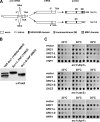
Alternative splicing of SRC1 results in two different spliced protein forms. (A) Schematic overview of pre-mRNA, mRNA, and protein products upon alternative splicing. Either a 126- or a 130-nt intron can be excised by using two alternative 5′ splice sites. In the latter case, a frame shift results in an earlier stop codon and, therefore, in a shorter protein with a different amino acid sequence at the C terminus compared with Src1-L. Conserved domains (HEH/LEM and MSC) and transmembrane domains (M) are indicated. Numbers represent amino acid residues. (B) Whole cell lysates of N- (TAP-Src1) and C-terminal TAP-tagged Src1-L or Src1-S were analyzed by SDS-PAGE followed by Western blotting using anti-ProtA antibodies. (C) Genetic relationship of SRC1 splice variants with TREX–THO and TREX-2 components. The double-disruption strains were transformed with empty vector, GFP-Src1 splice variants, and the respective TREX component. Transformants were spotted in 10-fold serial dilutions on 5-FOA–containing plates for 5 d at the indicated temperatures.
Figure 3.
Figure 3.
Src1-L and Src1-S are integral membrane proteins. (A) Kyte-Doolittle hydropathy analysis (with a 19–amino acid window size; Kyte and Doolittle, 1982) revealed two putative hydrophobic transmembrane-spanning regions (M1 and M2) for Src1-L but only one region with significant transmembrane potential for Src1-S. The hydrophobicity scores are plotted against the window number. (B) Crude membrane fractions from cells expressing Src1-L–TAP or Src1-S–TAP were extracted using four different conditions: 150 mM NaCl, 1 M NaCl, 1% Triton X-100, or pH 11.5. Equivalent amounts of fractions from lysate (L), membrane (P1), soluble (S1), and, after ultracentrifugation, insoluble pellet (P) and supernatant (S) were analyzed by SDS-PAGE followed by Western blotting with antibodies against ProtA, Vam3, and Nsp1. Western blotting against Vam3 and Nsp1 is only shown for Src1-S–TAP (top). Extraction profile of src1Δ cells harboring intron-containing constructs of ProtA-Src1 full length and deletion of the first (Src1ΔM1) or second (Src1ΔM2) hydrophobic stretch. Western blots were probed with an anti-ProtA antibody (bottom).
Figure 4.
Figure 4.
Src1-L is a double-pass integral membrane protein, and Src1-S is a single-pass integral membrane protein. (A) The six possible topological orientations for Src1-L and the two for Src1-S are shown. N, N terminus; C, C terminus; INM, inner nuclear membrane; ONM, outer nuclear membrane. Experimentally determined topologies are boxed in red. (B) Schematic illustration of the TEV-based method to determine the topology of Src1 membrane insertion (left). Under inducing conditions, the TEV protease is expressed and cleaves at TEV cleavage sites when accessible at the nuclear side. TEV-CS, TEV cleavage site. Cells expressing N- (TAP-Src1) or C-terminal TAP-tagged Src1 (Src1-L–TAP and Src1-S–TAP) or src1Δ cells containing C-terminal TAP-tagged Src1-L–ΔM2 were grown either under noninducing (−) or inducing (+) conditions (right). Whole cell extracts were analyzed by Western blotting using anti-ProtA, anti-myc (TEV protease), and anti-Arc1 antibodies (loading control).
Figure 5.
Figure 5.
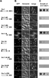
Src1 domain analysis. (A) Schematic representation of Src1 deletion constructs. (B) Fluorescence microscopy of src1Δ cells harboring the indicated GFP-tagged Src1 full-length and truncation constructs. (C) The double-disruption strain src1Δthp1Δ carrying plasmid-borne THP1 was transformed with the indicated constructs, and cells were spotted in 10-fold serial dilutions on 5-FOA–containing plates and incubated for 5 d at 30°C. Only cDNA-based constructs of Src1-L are shown.
Figure 6.
Figure 6.
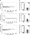
Analysis of gene expression in the src1Δ mutant. (A and B) Relative increases (src1/wt, when src1 > wt) or decreases (wt/src1, when src1 < wt) of gene expression were plotted versus their distance to the closest telomere (A) or centromere (B). A sliding window of 100 genes was used. The average of the 100 genes was used for the y axis, and the distance of the central gene in the window was used for the x axis. The bottom panel of A is an expanded view of the top graph showing the ∼27-kb region close to the telomeres, which exhibits a misregulation in the src1 deletion strain. (C) Expression levels of PHO mRNAs in wt and src1Δ cells. Total RNA of wt and src1Δ cells grown in HP and LP was prepared, and cDNA was analyzed by quantitative RT-PCR using specific primers for PHO11, PHO12, and PHO84. Each gene was assayed in triplicates. The mRNA levels of wt HP expression are set as one. One representative dataset of five times independently isolated RNA is shown. Error bars represent SD.
Figure 7.
Figure 7.
In vivo localization of PHO gene loci. 3D localization of TetO/TetR-GFP–labeled PHO11, PHO12, PHO84, and GIS1 loci in wt or src1Δ cells expressing GFP-Nup49 was determined after microscopy acquisition in vivo (see Materials and methods). For each strain, positions of the loci obtained from N nuclei are plotted in radial projection on the same graph (as previously described in Cabal et al., 2006). Hot colors indicate high density of loci, and cold colors indicate low density (the color scale is indicated on the bottom right). Average outlines of the nuclear envelope (orange circles) are also shown.
Figure 8.
Figure 8.
Genome-wide distribution maps of Src1. ChIP-on-chip analyses were performed to identify the global targets of Src1-L across the S. cerevisiae genome. Relative enrichments of ProtA-Src1 or ProtA as a control are plotted in alignment with the map of each chromosome. For simplicity, only four representative chromosomes are shown. Localization of PHO11 and PHO12, the rDNA locus, and the two mating-type loci is shown. cen, centromere; HML/HMR, mating-type loci; tel, telomere.
Figure 9.
Figure 9.
src1Δ cells are not affected in telomere silencing, but in combination with the yku70Δ mutation, they are synergistically impaired. Deletions of SRC1, YKU70, or double deletion in a telVIIL∷URA3 strain were spotted in 10-fold serial dilutions on 5-FOA–containing plates and grown for 5 d at 23°C. Note that growth reflects telomeric silencing and reduced growth derepression of silencing. Synthetic dextrose complete without 5-FOA was used as a plating control.

Similar articles

Cited by

References

    1. Akhtar, A., and S.M. Gasser. 2007. The nuclear envelope and transcriptional control. Nat. Rev. Genet. 8:507–517. - PubMed
    1. Alberola, T.M., J. García-Martínez, O. Antúnez, L. Viladevall, A. Barcelo, J. Ariño, and J.E. Pérez-Ortín. 2004. A new set of DNA macrochips for the yeast Saccharomyces cerevisiae: features and uses. Int. Microbiol. 7:199–206. - PubMed
    1. Askree, S.H., T. Yehuda, S. Smolikov, R. Gurevich, J. Hawk, C. Coker, A. Krauskopf, M. Kupiec, and M.J. McEachern. 2004. A genome-wide screen for Saccharomyces cerevisiae deletion mutants that affect telomere length. Proc. Natl. Acad. Sci. USA. 101:8658–8663. - PMC - PubMed
    1. Bernstein, B.E., E.L. Humphrey, R.L. Erlich, R. Schneider, P. Bouman, J.S. Liu, T. Kouzarides, and S.L. Schreiber. 2002. Methylation of histone H3 Lys 4 in coding regions of active genes. Proc. Natl. Acad. Sci. USA. 99:8695–8700. - PMC - PubMed
    1. Brachner, A., S. Reipert, R. Foisner, and J. Gotzmann. 2005. LEM2 is a novel MAN1-related inner nuclear membrane protein associated with A-type lamins. J. Cell Sci. 118:5797–5810. - PubMed

Publication types

MeSH terms