. 2008 Nov 20;27(54):6791-805.
doi: 10.1038/onc.2008.291. Epub 2008 Sep 1.

Smad7 is inactivated through a direct physical interaction with the LIM protein Hic-5/ARA55

Affiliations

Smad7 is inactivated through a direct physical interaction with the LIM protein Hic-5/ARA55

H Wang et al. Oncogene. .

Erratum in

Abstract

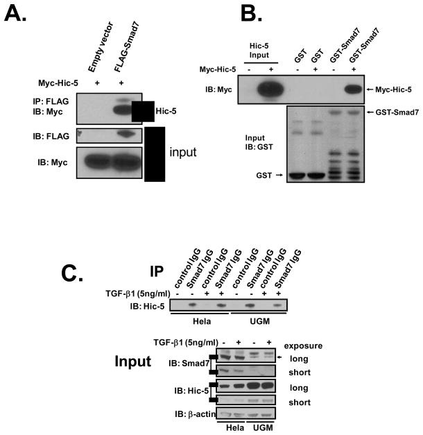
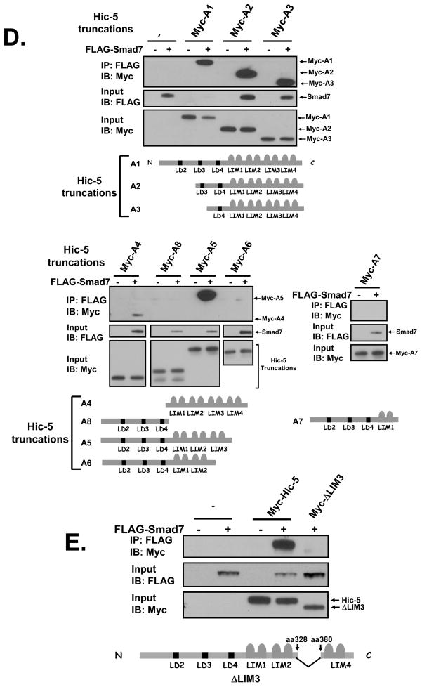
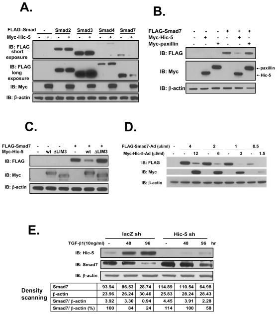
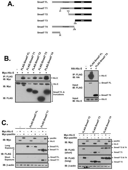
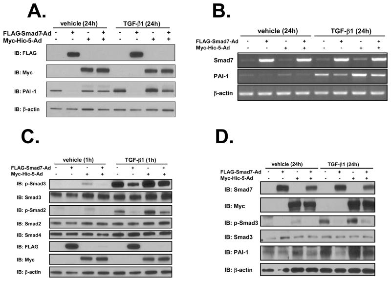
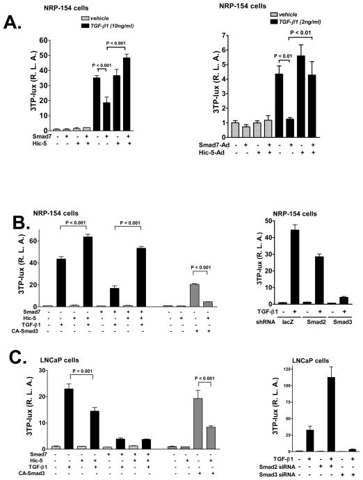
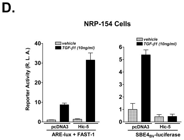
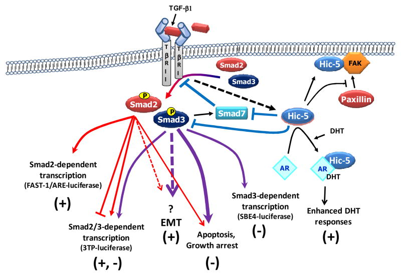
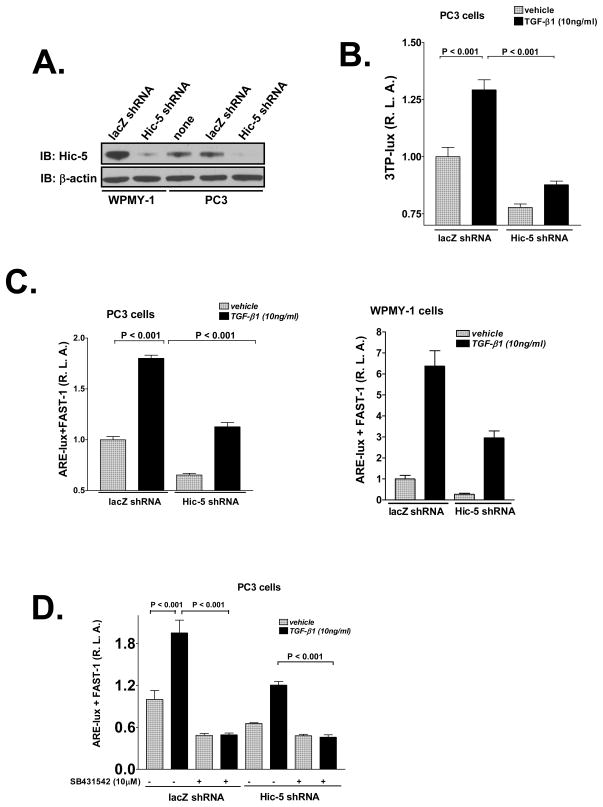
We recently reported that hydrogen peroxide-inducible clone-5 (Hic-5, also named androgen receptor-associated protein 55) can bind to the transforming growth factor-beta (TGF-beta)-signaling regulator Smad3, thereby inhibiting certain Smad3-dependent TGF-beta responses. We now show that Hic-5 can also control TGF-beta responses through an alternative mechanism involving Smad7, a key negative regulator of TGF-beta signaling. Hic-5 binds directly to Smad7. This interaction requires the LIM3 domain of Hic-5, and enhances TGF-beta signaling through causing loss of Smad7 protein but not mRNA. Enforced expression of Hic-5 reverses the ability of Smad7 to suppress TGF-beta-induced phosphorylation of Smads 2 and 3 and activation of the plasminogen activator inhibitor-1 promoter (in NRP-154 and PC3 prostate carcinoma and WPMY-1 prostate myofibroblast cell lines). Lentiviral-mediated small-hairpin RNA silencing of endogenous Hic-5 reduced TGF-beta responses in PC3 and WPMY-1 cells. Further work suggests that the level of Smad7 is modulated by its physical interaction with Hic-5 and targeted to a degradation pathway not likely to be proteasomal. Our findings support that Hic-5 functions as a cell-type-specific activator of TGF-beta signaling through its ability to physically interact with and neutralize Smad7.


Figures

Figure 1
Figure 1. Binding of Hic-5 to Smad7 through Hic-5 LIM3 domain
A, Interaction between Hic-5 and Smad7. HEK293 cells were co-transfected with 1 μg/well of Myc-Hic-5 and 1μg/well of FLAG-Smad7 or the empty vector. Cell lysates were immunoprecipitated with anti-FLAG M2 monoclonal antibody, and both the immunoprecipitated proteins and the input cells lysates were immunoblotted with anti-Myc antibody A-14. B, the binding of Smad7 to Hic-5 is direct. In vitro transcribed and translated Myc-Hic-5 was incubated with GST or GST-Smad7 fusion protein overnight at 4°C in the presence of glutathione-Sepharose 4B beads. Elution of GST from the beads by treatment (50° C, 5 min) with SDS-PAGE sample loading buffer released both GST-Smad7 and Myc-Hic-5 as detected following immunoblotting with anti-GST and anti-Myc A-14 antibodies, respectively. C, Hela or rat urogenital sinus mesenchymal (UGM) cells were plated in 100-mm dishes with 5% FBS/DMEM/F-12 medium. Twenty-four hours before harvesting, TGF-β1 (5 ng/ml) or vehicle was added to cells. Cells were lysed in cold RIPA buffer and then the endogenous Smad7 was immunoprecipitated with control rabbit IgG or anti-Smad7 rabbit IgG. The resultant eluates were immunoblotted with anti-Hic-5 mouse antibody. D and E, Hic-5 LIM3 domain is the binding region. HEK293 cells were co-transfected with 1 μg/well of full-length Myc-Hic-5 (Hic-5 A1), Myc-Hic-5 truncation vectors (Hic-5 A2 – Hic-5 A8, see map in figure), or Myc-Hic-5 without the LIM3 domain (Hic-5ΔLIM3) and 1 μg/well of FLAG-Smad7 or the empty vector. Immunoprecipitation and immunoblotting were completed in the same way as described in A. All of the data shown are representative of two to three independent experiments.
Figure 2
Figure 2. Binding of Hic-5 to Smad7 leads to Smad7 protein loss
A, Hic-5 selectively causes Smad7 protein loss. HEK293 cells were co-transfected with 1 μg/well of vectors bearing FLAG-Smad cDNA fragments and 1 μg/well of Myc-Hic-5 or the empty vector. The same amount of cell lysate was immunoblotted to detect tagged proteins. B and C, Binding is necessary for Smad7 protein reduction by Hic-5. HEK293 (B) or HEK293T (C) cells were co-transfected with 1 μg/well of the empty vector or Myc-tagged plasmids (Hic-5, paxillin or Hic-5 ΔLIM3) and 1 μg/well of FLAG-Smad7. Tagged proteins were detected as described in A. D, Downregulation of Smad7 protein by Hic-5 in NRP-154 cells. NRP-154 cells were co-infected with different doses (v/v dilution) of the adenoviruses Myc-Hic-5-AdMax (Ad) and FLAG-Smad7-Ad overnight. After 36 h, cells were harvested for immunoblotting to detect the expression of exogenous proteins. E, Silencing Hic-5 in DU145 cells. DU145 cells infected with lentivirus bearing lacZ shRNA or Hic-5 shRNA were treated with vehicle or TGF-β1 (10 ng/ml, recombinant human TGF-β1, R&D Systems, Inc.) for 48 h or 96 h. Band intensities were evaluated with Quantity One 4.5.1 (Bio-Rad). Cell lysates were immunoblotted for Hic-5 and Smad7, respectively. All of the data shown are representative of three independent experiments.
Figure 3
Figure 3. Interaction of Smad7 truncations with Hic-5
A, Smad7 truncation constructs. B, Binding of Smad7 truncations to Hic-5. HEK293 cells were co-transfected with 1 μg/well of Myc-Hic-5 and 1 μg/well of FLAG-Smad7 truncations or the empty vector. Cell lysates were immunoprecipitated with anti-FLAG M2 monoclonal antibody, and both the immunoprecipitated proteins and input lysates were immunoblotted with anti-Myc antibody A-14. C, Effects of Hic-5 on Smad7 truncation protein levels. HEK293 cells were co-transfected with 2 μg/well vectors bearing 1 μg Myc-Hic-5 and 1 μg of each of the FLAG-Smad7 truncations or the empty vector. Cell lysate was immunoblotted to detect tagged proteins. All of the data shown are representative of two independent experiments.
Figure 4
Figure 4. Effects of Hic-5-Smad7 interaction on TGF-β-induced R-Smad activation and PAI-1 expression
A and D, NRP-154 cells (A) or PC3 cells (D) were co-infected with ±FLAG-Smad7-Ad (1:1000 v/v dilution) and ±Myc-Hic-5-Ad (3:1000 v/v dilution) overnight. Then cells were added with vehicle or 10 ng/ml TGF-β1. Twenty-four h later, cells were harvested for immunoblotting to detect protein level of Hic-5 or Smad7 and endogenous protein level of PAI-1 and β-actin (loading control). B, NRP-154 cells were treated in the same way as described in A but mRNA levels of Smad7 and endogenous PAI-1 were measured. C, NRP-154 cells were treated as described in A. However, TGF-β1 (10 ng/ml) was added 1 h before harvesting. All of the data shown are representative of three independent experiments.
Figure 5
Figure 5. Inactivation of Smad7 by Hic-5
A, Left panel, NRP-154 cells were co-transfected (calcium phosphate method (Wang et al., 2005)) with 0.2 μg/well of 3TP-lux reporter, 12.5 ng/well of pRL-CMV internal control, ±0.1 μg/well of FLAG-Smad7 and ±0.7 μg/well of Myc-Hic-5. The next day, cells were treated with 10 ng/ml TGF-β1 or vehicle for 24 h before harvesting. Right panel, NRP-154 cells were infected with FLAG-Smad7-Ad (1:1000 v/v dilution) and Myc-Hic-5-Ad (3:1000 v/v dilution) constructs and then were transfected with the same amount of 3TP-lux reporter and pRL-CMV internal control plasmid as used in A. The next day, 2 ng/ml TGF-β1 or vehicle was used to treat cells for 24 h before harvesting. B, NRP-154 cells were co-transfected with the same amount of various plasmids as described in A, except that ±0.65 μg/well of Myc-Hic-5 was used in this experiment. The next day, 10 ng/ml TGF-β1 or vehicle was added and luciferase activity was measured 24 h later. CA-Smad3 (0.1 μg/well) was used as an inducer in a parallel experiment, where cells were harvested 24 h after transfection. Smad2 and Smad3 were silenced with shRNA according to our recent published protocol (Yang et al., 2008). C, LNCaP cells were treated in the same way as described in B. Smad2 and Smad3 were silenced with siRNA (Supplementary Figure 5) according to another publication form our laboratory (Song et al., 2006b). D, NRP-154 cells were co-transfected with 0.2 μg/well of reporter (ARE-lux/FAST-1 or SBE4BV-luciferase), 20 ng/well of pRL-CMV internal control and ±0.8 μg/well of Myc-Hic-5. The next day, cells were treated with 10 ng/ml TGF-β1 or vehicle for 24 h before harvesting for luciferase test. In all reporter assays, dual luciferase tests were performed as described before (Wang et al., 2005). Luciferase values represent the average of triplicate determinations ±SEM. All of the data shown are representative of three independent experiments.
Figure 6
Figure 6. Signal transduction diagram depicting how Hic-5 affects TGF-β signaling
Hic-5, which is induced by TGF-β through a mechanism that is yet to be resolved, binds to and inactivates both Smad3 and Smad7. Inactivation of Smad7 through its degradation relieves the inhibitory action of Smad7 on TGF-β receptors, which leads to phosphorylation and thus activation of Smads 2 and 3. Depletion of Smad7 frees up Hic-5 and is expected to enable further binding to and suppression of Smad3. We thus propose that the overall effect of Hic-5 on TGF-β signaling is inhibition of Smad3-dependent pathway and enhancement of Smad2-dependent or Smad-independent pathway.
Figure 7
Figure 7. Role of Endogenous Hic-5 on TGF-β responses
A, Silencing Hic-5 in WPMY-1 and PC3 cells. WPMY-1 or PC3 cells were infected with lentiviral lacZ shRNA or Hic-5 shRNA construct overnight and then maintained in culture medium. After 1 day, cells were harvested and endogenous Hic-5 protein was detected with anti-Hic-5 antibody. B, PC3 cells bearing lacZ shRNA or Hic-5 shRNA were transfected with 0.2 μg/well of 3TP-lux reporter and 20 ng/well of pRL-CMV internal control. The next day, cells were treated with 10 ng/ml TGF-β1 or vehicle for 24 h and then harvested for luciferase test. C, PC3 or WPMY-1 cells bearing lacZ shRNA or Hic-5 shRNA were treated as described in B except that ARE-lux co-expressed with FAST-1 was used as the reporter. D, PC3 cells bearing lacZ shRNA or Hic-5 shRNA were treated as described in C. However, after transfection, cells were added with 10 μM TβRI kinase inhibitor SB431542 or vehicle. Luciferase values in B, C and D represent the average of triplicate determinations ±SEM. All of the data shown are representative of two to three independent experiments.

Similar articles

Cited by

References

    1. Chen X, Weisberg E, Fridmacher V, Watanabe M, Naco G, Whitman M. Smad4 and FAST-1 in the assembly of activin-responsive factor. Nature. 1997;389:85–9. - PubMed
    1. Chipuk JE, Cornelius SC, Pultz NJ, Jorgensen JS, Bonham MJ, Kim SJ, et al. The androgen receptor represses transforming growth factor-beta signaling through interaction with Smad3. J Biol Chem. 2002a;277:1240–8. - PubMed
    1. Chipuk JE, Stewart LV, Ranieri A, Song K, Danielpour D. Identification and characterization of a novel rat ov-serpin family member, trespin. J Biol Chem. 2002b;277:26412–21. - PubMed
    1. Dabiri G, Tumbarello DA, Turner CE, Van De Water L. Hic-5 Promotes the Hypertrophic Scar Myofibroblast Phenotype by Regulating the TGF-beta1 Autocrine Loop. J Invest Dermatol 2008 - PMC - PubMed
    1. Danielpour D. Functions and regulation of transforming growth factor-beta (TGF-beta) in the prostate. Eur J Cancer. 2005;41:846–57. - PubMed

Publication types

MeSH terms

Substances