

Brain mu-opioid receptor binding: relationship to relapse to cocaine use after monitored abstinence
- PMID: 18762918
- PMCID: PMC2575005
- DOI: 10.1007/s00213-008-1225-5
Brain mu-opioid receptor binding: relationship to relapse to cocaine use after monitored abstinence
Abstract
Rationale: Cocaine users have increased regional brain mu-opioid receptor (mOR) binding which correlates with cocaine craving. The relationship of mOR binding to relapse is unknown.
Objective: To evaluate regional brain mOR binding as a predictor of relapse to cocaine use is the objective of the study.
Materials and methods: Fifteen nontreatment-seeking, adult cocaine users were housed on a closed research ward for 12 weeks of monitored abstinence and then followed for up to 1 year after discharge. Regional brain mOR binding was measured after 1 and 12 weeks using positron emission tomography (PET) with [11C]carfentanil (a selective mOR agonist). Time to first cocaine use (lapse) and to first two consecutive days of cocaine use (relapse) after discharge was based on self-report and urine toxicology.
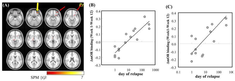
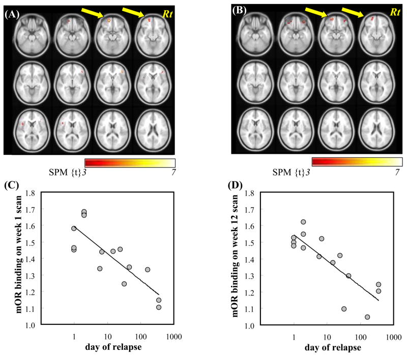
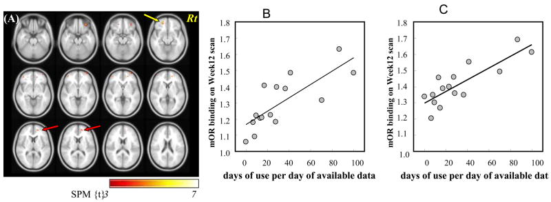
Results: A shorter interval before relapse was associated with increased mOR binding in frontal and temporal cortical regions at 1 and 12 weeks of abstinence (Ps < 0.001) and with a lesser decrease in binding between 1 and 12 weeks (Ps < 0.0008). There were significant positive correlations between mOR binding at 12 weeks and percent days of cocaine use during first month after relapse (Ps < 0.002). In multiple linear regression analysis, mOR binding contributed significantly to the prediction of time to relapse (R2= 0.79, P < 0.001), even after accounting for clinical variables.
Conclusions: Increased brain mOR binding in frontal and temporal cortical regions is a significant independent predictor of time to relapse to cocaine use, suggesting an important role for the brain endogenous opioid system in cocaine addiction.
Figures
Similar articles
-
Brain mu-opioid receptor binding predicts treatment outcome in cocaine-abusing outpatients.Biol Psychiatry. 2010 Oct 15;68(8):697-703. doi: 10.1016/j.biopsych.2010.05.003. Epub 2010 Jul 1. Biol Psychiatry. 2010. PMID: 20579973 Free PMC article.
-
Imaging brain mu-opioid receptors in abstinent cocaine users: time course and relation to cocaine craving.Biol Psychiatry. 2005 Jun 15;57(12):1573-82. doi: 10.1016/j.biopsych.2005.02.026. Biol Psychiatry. 2005. PMID: 15953495
-
Differential response to IV carfentanil in chronic cocaine users and healthy controls.Addict Biol. 2012 Jan;17(1):149-55. doi: 10.1111/j.1369-1600.2010.00256.x. Epub 2010 Nov 4. Addict Biol. 2012. PMID: 21054687 Free PMC article.
-
PET imaging of dopamine D2 receptors in monkey models of cocaine abuse: genetic predisposition versus environmental modulation.Am J Psychiatry. 2005 Aug;162(8):1473-82. doi: 10.1176/appi.ajp.162.8.1473. Am J Psychiatry. 2005. PMID: 16055768 Review.
-
Dopamine and opioid systems adaptation in alcoholism revisited: Convergent evidence from positron emission tomography and postmortem studies.Neurosci Biobehav Rev. 2019 Nov;106:141-164. doi: 10.1016/j.neubiorev.2018.09.010. Epub 2018 Sep 19. Neurosci Biobehav Rev. 2019. PMID: 30243576
Cited by
-
Blunted Endogenous Opioid Release Following an Oral Amphetamine Challenge in Pathological Gamblers.Neuropsychopharmacology. 2016 Jun;41(7):1742-50. doi: 10.1038/npp.2015.340. Epub 2015 Nov 10. Neuropsychopharmacology. 2016. PMID: 26552847 Free PMC article.
-
A Survey of Molecular Imaging of Opioid Receptors.Molecules. 2019 Nov 19;24(22):4190. doi: 10.3390/molecules24224190. Molecules. 2019. PMID: 31752279 Free PMC article. Review.
-
Fischer 344 and Lewis Rat Strains as a Model of Genetic Vulnerability to Drug Addiction.Front Neurosci. 2016 Feb 9;10:13. doi: 10.3389/fnins.2016.00013. eCollection 2016. Front Neurosci. 2016. PMID: 26903787 Free PMC article. Review.
-
Toward biomarkers of the addicted human brain: Using neuroimaging to predict relapse and sustained abstinence in substance use disorder.Prog Neuropsychopharmacol Biol Psychiatry. 2018 Jan 3;80(Pt B):143-154. doi: 10.1016/j.pnpbp.2017.03.003. Epub 2017 Mar 18. Prog Neuropsychopharmacol Biol Psychiatry. 2018. PMID: 28322982 Free PMC article. Review.
-
Role of mu- and delta-opioid receptors in the nucleus accumbens in cocaine-seeking behavior.Neuropsychopharmacology. 2009 Jul;34(8):1946-57. doi: 10.1038/npp.2009.28. Epub 2009 Mar 11. Neuropsychopharmacology. 2009. PMID: 19279569 Free PMC article.
References
-
- American Psychiatric Association. Diagnostic and Statistical Manual of Mental Disorders. 4. American Psychiatric Association; Washington, D.C.: 1994.
-
- Azaryan AV, Clock BJ, Cox BM. Mu opioid receptor mRNA in nucleus accumbens is elevated following dopamine receptor activation. Neurochem Res. 1996;21:1411–1415. - PubMed
-
- Azaryan AV, Coughlin LJ, Buzas B, Clock BJ, Cox BM. Effect of chronic cocaine treatment on mu- and delta-opioid receptor mRNA levels in dopaminergically innervated brain regions. J Neurochem. 1996;66:443–448. - PubMed
-
- Bauer LO. Frontal P300 decrements, childhood conduct disorder, family history, and the prediction of relapse among abstinent cocaine abusers. Drug Alcohol Depend. 1997;44:1–10. - PubMed
-
- Brady KT, Sonne SC, Malcolm RJ, Randall CL, Dansky BS, Simpson K, Roberts JS, Brondino M. Carbamazepine in the treatment of cocaine dependence: subtyping by affective disorder. Exp Clin Psychopharmacol. 2002;10:276–285. - PubMed
Publication types
MeSH terms
Substances
Grants and funding
LinkOut - more resources
Full Text Sources
Medical
Research Materials
