

Evaluation of the femoral midshaft in children with cerebral palsy using magnetic resonance imaging
- PMID: 18763012
- PMCID: PMC5992489
- DOI: 10.1007/s00198-008-0718-8
Evaluation of the femoral midshaft in children with cerebral palsy using magnetic resonance imaging
Abstract
Summary: Magnetic resonance imaging was used to show that children with quadriplegic cerebral palsy and unable to ambulate independently compared to typically developing children have a remarkably underdeveloped femoral midshaft as indicated by a very thin diameter, a very thin cortical wall, and very low strength estimates.
Introduction: The femoral shaft is very susceptible to fracture in children with quadriplegic cerebral palsy (QCP); however, its structure and strength have not been evaluated.
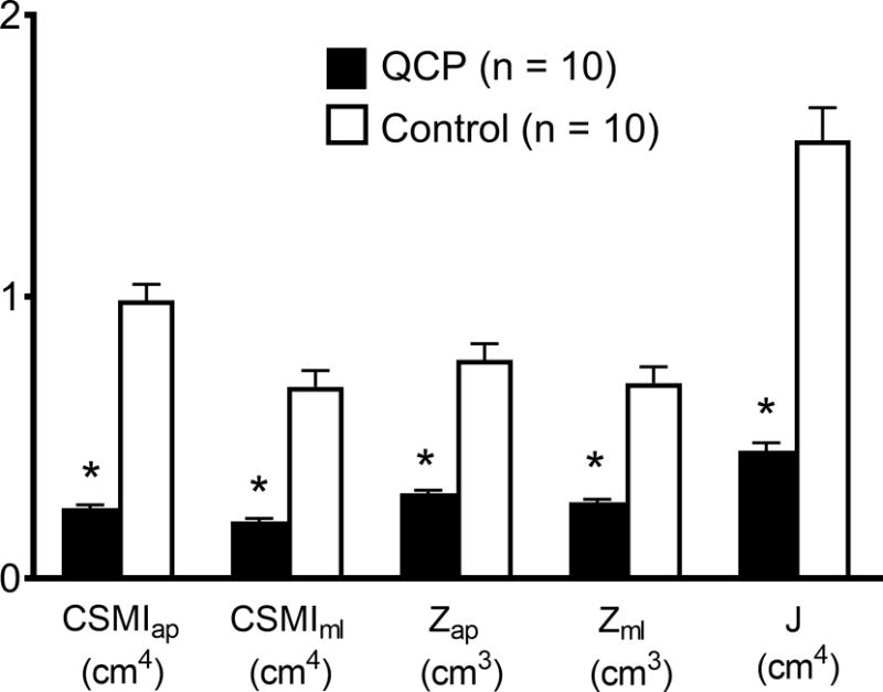
Methods: The volume and width of the middle third of the femur (midfemur) and its cortical wall and medullary cavity were assessed in children with QCP and unable to ambulate independently and typically developing children (n = 10/group) using magnetic resonance imaging (MRI). Estimates of cross-sectional moment of inertia (CSMI), section modulus (Z), and polar moment of inertia (J) were also determined.
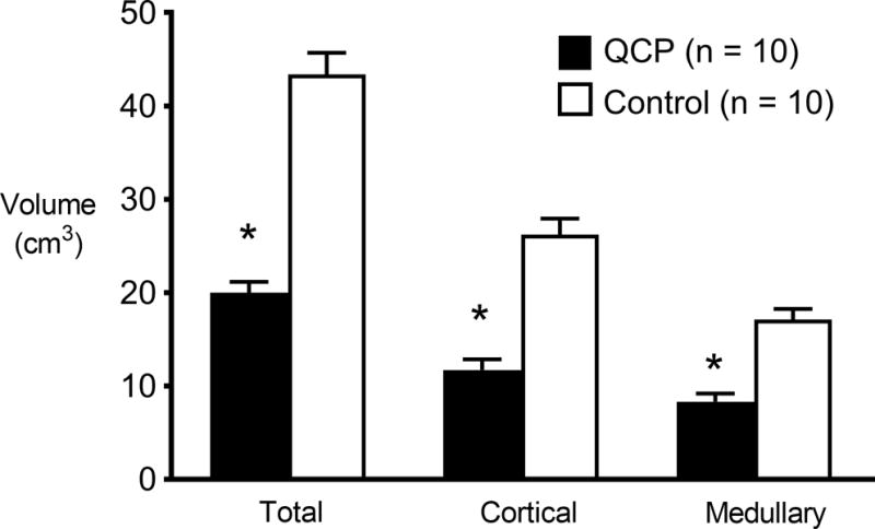
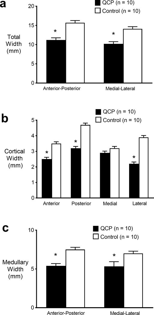
Results: Total volume of the midfemur and volume of its cortical wall and medullary cavity were substantially lower in children with QCP than controls (51-55%; p < 0.001). In addition, the total midfemur, its medullary cavity and the anterior, posterior, and lateral sections of its cortical wall were thinner (27-43%) in children with QCP (p < 0.001). The midfemur in children with QCP also had remarkably lower CSMI, Z, and J (60-71%; p < 0.001).
Conclusions: Children with QCP who lack the ability to ambulate independently have midfemurs that are very thin with very thin cortical walls and very low estimated strength. The disparity can be detected using MRI.
Conflict of interest statement
Conflicts of Interest: The authors declare that they have no conflict of interest or disclosures.
Figures
References
-
- McIvor WC, Samilson RL. Fractures in patients with cerebral palsy. J Bone Joint Surg Am. 1966;48:858–866. - PubMed
-
- Presedo A, Dabney KW, Miller F. Fractures in patients with cerebral palsy. J Pediatr Orthop. 2007;27:147–153. - PubMed
-
- Henderson RC, Lark RK, Gurka MJ, Worley G, Fung EB, Conaway M, Stallings VA, Stevenson RD. Bone density and metabolism in children and adolescents with moderate to severe cerebral palsy. Pediatrics. 2002;110:e5. - PubMed
-
- Modlesky CM, Subramanian P, Miller F. Underdeveloped trabecular bone microarchitecture is detected in children with cerebral palsy using high-resolution magnetic resonance imaging. Osteoporos Int. 2008;19:169–176. - PubMed
-
- Ciarelli TE, Fyhrie DP, Schaffler MB, Goldstein SA. Variations in three-dimensional cancellous bone architecture of the proximal femur in female hip fractures and in controls. J Bone Miner Res. 2000;15:32–40. - PubMed
