

Effects of different types of palatal lateral excisions on growth and development of maxilla and dental arch
- PMID: 18763314
- PMCID: PMC2491694
- DOI: 10.1631/jzus.B0720015
Effects of different types of palatal lateral excisions on growth and development of maxilla and dental arch
Abstract
Objective: This study aimed to explore the effects of different types of palatal lateral excisions on the growth and development of the maxilla and dental arch, and to investigate the underlying mechanisms.
Methods: A total of 112 3-week-old Sprague-Dawley (SD) male rats were randomly divided into a control and 3 experimental groups: the mucoperiosteal denudation group, the mucosal flap excision group, and the periosteum excision group. In the experimental groups, bilateral mucoperiosteal, mucosal flap and periosteum were excised respectively in the lateral one half of the palate. Four rats in each group were randomly chosen for sacrifice every two weeks. The maxilla was dissected following the excision. The widths of the maxilla and dental arch were measured and the histological phenomena were investigated at different phases. At the same time, 12 animals in each group were sequentially injected with calcein every two weeks. Three animals in each group, whose fluorescent labeling was used, were sacrificed for investigating bone formation at Week 8 following injection.
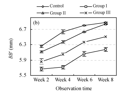
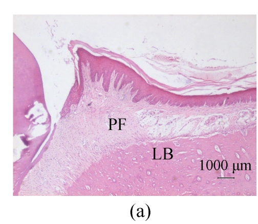
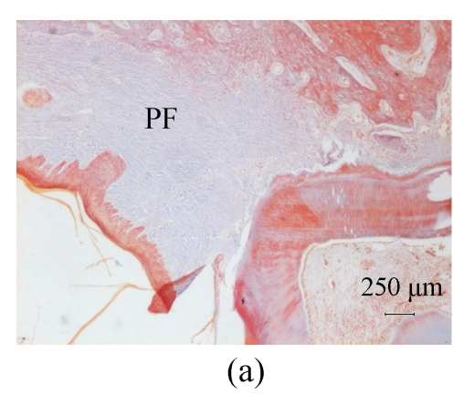
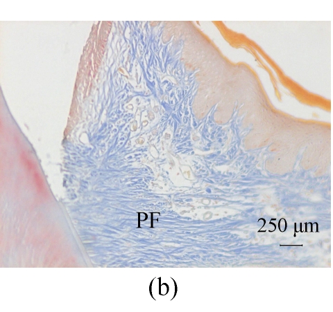
Results: (1) Each experimental group presented the constriction of the maxilla and dental arch. The upper first molars in the experimental groups inclined medially. The mucoperiosteal denudation group showed the largest degree of effect followed by the periosteum excision group. The indices of the mucosal flap excision group, which retained the structures of the periosteum layer, had the most approximate values to the control group; (2) Different histological changes among the experimental groups were detected. The fibers penetrated into the palatal bone as Sharpey's fibers in the mucoperiosteal denudation group. The pattern of bone deposition was the bundle type. Sharpey's fibers were not found in the mucosal flap and periosteum excision groups and the depositions of palatal bone were the lamellar type as those in the control group; (3) The rates of bone deposition in the experimental groups decreased compared with the control group. The rates in different phases were the most approximate values to those of the control group in the mucosal flap excision group, which has the same structure of periosteum as the control group.
Conclusion: There were different effects on the growth and development of the maxilla and dental arch in different types of palatal lateral excisions. Periosteum is important for bone formation and deposition pattern. The prevention of Sharpey's fibers forming and attaching to the palatine can effectively avert the following malformation.
Figures


Similar articles
-
Roles of different areas of palatine bone denudation on growth and development of the maxilla and dental arch: an experimental study.J Craniofac Surg. 2007 Mar;18(2):391-8. doi: 10.1097/01.scs.0000235109.15860.f7. J Craniofac Surg. 2007. PMID: 17414291
-
Effects of palatine bone denudation repair with periosteal graft on maxillary growth: an experimental study in rats.J Craniomaxillofac Surg. 2014 Jan;42(1):e1-7. doi: 10.1016/j.jcms.2013.02.008. Epub 2013 Mar 20. J Craniomaxillofac Surg. 2014. PMID: 23523011
-
Constriction of the maxillary dental arch by mucoperiosteal denudation of the palate.Cleft Palate Craniofac J. 2002 Jul;39(4):425-31. doi: 10.1597/1545-1569_2002_039_0425_cotmda_2.0.co_2. Cleft Palate Craniofac J. 2002. PMID: 12071790
-
Maxillary growth following atelocollagen implantation on mucoperiosteal denudation of the palatal process in young rabbits: implications for clinical cleft palate repair.Cleft Palate Craniofac J. 1997 Jul;34(4):297-308. doi: 10.1597/1545-1569_1997_034_0297_mgfaio_2.3.co_2. Cleft Palate Craniofac J. 1997. PMID: 9257020
-
Effect of timing of palatal repair on the transverse development of maxillary alveolar arch in complete-cleft cases.J Indian Soc Pedod Prev Dent. 1991 Mar;8(1):15-8. J Indian Soc Pedod Prev Dent. 1991. PMID: 2056341 Review.
References
-
- Diehl H, Ellingboe JL. Indicator for titration of calcium in presence of magnesium using disodium dihydrogen ethylenediamine tetraacetate. Anal Chem. 1956;28(5):882–884. doi: 10.1021/ac60113a030. - DOI
-
- Graber TM. A ceghalometric analysis of the developmental pattern and facial morphology in cleft palate. Angle Orthod. 1949;19(2):91–100.
MeSH terms
LinkOut - more resources
Full Text Sources
