. 2008 Sep 2:3:12.
doi: 10.1186/1750-1326-3-12.

Evidence that the Amyloid beta Precursor Protein-intracellular domain lowers the stress threshold of neurons and has a "regulated" transcriptional role

Affiliations

Evidence that the Amyloid beta Precursor Protein-intracellular domain lowers the stress threshold of neurons and has a "regulated" transcriptional role

Luca Giliberto et al. Mol Neurodegener. .

Abstract

Background: Regulated intramembrane proteolysis of the beta-amyloid precursor protein by the gamma-secretase yields two peptides. One, amyloid-beta, is the major component of the amyloid plaques found in Alzheimer's disease patients. The other, APP IntraCellular Domain, has been involved in regulation of apoptosis, calcium flux and gene transcription. To date, a few potential target genes transcriptionally controlled by AID, alone or complexed with Fe65/Tip60, have been described. Although the reports are controversial: these include KAI1, Neprilysin, p53, EGFR, LRP and APP itself. Furthermore, p53 has been implicated in AID mediated susceptibility to apoptosis. To extend these findings, and assess their in vivo relevance, we have analyzed the expression of the putative target genes and of the total brain basal transriptoma in transgenic mice expressing AID in the forebrain. Also, we have studied the susceptibility of primary neurons from such mice to stress and pro-apoptotic agents.

Results: We found that AID-target genes and the mouse brain basal transcriptoma are not influenced by transgenic expression of AID alone, in the absence of Fe65 over-expression. Also, experiments conducted on primary neurons from AID transgenic mice, suggest a role for AID in sensitizing these cells to toxic stimuli. Overall, these findings hint that a role for AID, in regulating gene transcription, could be induced by yet undefined, and possibly stressful, stimuli in vivo.

Conclusion: Overall, these data suggest that the release of the APP intracellular domain may modulate the sensitivity of neuronal cells to toxic stimuli, and that a transcriptional role of AID could be inscribed in signaling pathways thatare not activated in basal conditions.


Figures

Figure 1
Figure 1
Characterization of AID transgenic mice. A Schematic representation of the transgenic AID constructs (fragments are not depicted in scale). The location of the PCR primers a and b used for genotyping is shown. B PCR of 63 pups (18 are shown here) revealed that 9 of them (three are shown here, number 1, 4 and 15) had integrated the AID transgene. In the same PCR tube, β-actin was amplified to control for genomic DNA content. Vec. represents the control PCR performed using the transgenic vector as a template. C Real Time PCR showing the expression levels of the AID transgene in different lines. D Immunoprecipitation and western blot was conducted from a brain hemisphere of AIDtg and littermate (Con) mice. Mice lines that expressed the AID protein at the mRNA level, and had a detectable band at the western blot analysis were selected for further studies.
Figure 2
Figure 2
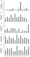
In vivo expression of candidate AID targets is not affected by transgenic AID expression, in adult mice. Real Time Quantitative PCR shows the relative expression of AIDtg protein, A, APP, B, NEP, C, KAI1, D, and p53, E in the forebrain and hippocampus of AIDtg and their littermate control mice. Values are relative to 100% value given arbitrarily to a littermate mouse. Experiments were conducted in triplicate loading, and the error bars represent standard deviations.
Figure 3
Figure 3
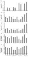
AID is already expressed at postnatal day nine, but does not influence the expression of the candidate genes. Real Time Quantitative PCR shows the relative expression of AIDtg protein A, APP, B, NEP, C, KAI1, D, and p53, E in the forebrain and hippocampus of AIDtg and their littermate control mice as early as 9 and 18 days post-natal. For APP, NEP, KAI1 and p53 expression, values are relative to 100% value given arbitrarily their day 9 and day 18 littermates. For AIDtg protein, 100% value was assigned to the first day 9 littermate mouse. Experiments were conducted in triplicate loading, and the error bars represent standard deviations.
Figure 4
Figure 4
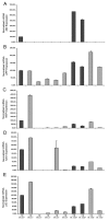
AIDtg expression in cultured fetal primary neurons does not change the relative expression of APP, NEP, KAI1 and p53. A AIDtg expression was confirmed by tail genotyping of fetuses (not shown) and by QPCR data on cultured neurons (dark bars). Expression of AID, A, APP, B, NEP, C, KAI1, D, and p53, E, is relative to the 100% value given arbitrarily to the first AIDtg mouse. Experiments were conducted in triplicate loading, and the error bars represent standard deviations. Cultures were harvested at DIV 14. Similar results were achieved from younger cultures (DIV 9, not shown) and in the AIDTg 59 line (not shown).
Figure 5
Figure 5
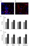
AIDtg expression in cultured fetal primary neurons increases their sensitivity to toxic and apoptotic stimuli. AIDTg line 59.4.4, at DIV 9. To verify the purity of the neuronal cultures, cells were stained with anti-NeuN (Blue) plus anti-GFAP (red), A, and NeuN (Blue) plus anti-MAP2 (red), B. C Toxicity of indicated stimulus was assessed by measuring LDH release. D Released LDH was weighed against WST-1 uptake in the LDH/WST-1 ratio, which confirms the trend of LDH release. C and D Values from toxic stimuli were weighed against values from untreated cells to express the increase of the indicators of cell damage. Average values refer to at least 3 AIDtg and 3 littermate mice; measurements were done on 6 separate wells of 96 well culture plate for each foetus' neurons. Similar results were obtained for AIDTg 57 line (not shown).

References

    1. Zheng H, Koo EH. Mol Neurodegener. 2006/08/26. Vol. 1. 2006. The amyloid precursor protein: beyond amyloid; p. 5. - DOI - PMC - PubMed
    1. Selkoe DJ, Podlisny MB. Deciphering the genetic basis of Alzheimer's disease. Annu Rev Genomics Hum Genet. 2002;3:67–99. doi: 10.1146/annurev.genom.3.022502.103022. - DOI - PubMed
    1. Passer B, Pellegrini L, Russo C, Siegel RM, Lenardo MJ, Schettini G, Bachmann M, Tabaton M, D'Adamio L. Generation of an apoptotic intracellular peptide by gamma-secretase cleavage of Alzheimer's amyloid beta protein precursor. J Alzheimers Dis. 2000;2:289–301. - PubMed
    1. Leissring MA, Murphy MP, Mead TR, Akbari Y, Sugarman MC, Jannatipour M, Anliker B, Muller U, Saftig P, De Strooper B, Wolfe MS, Golde TE, LaFerla FM. Proc Natl Acad Sci U S A. 2002/03/28. Vol. 99. 2002. A physiologic signaling role for the gamma -secretase-derived intracellular fragment of APP; pp. 4697–4702. - DOI - PMC - PubMed
    1. Cao X, Sudhof TC. A transcriptionally [correction of transcriptively] active complex of APP with Fe65 and histone acetyltransferase Tip60. Science. 2001;293:115–120. doi: 10.1126/science.1058783. - DOI - PubMed