Comparative Study
. 2008 Sep 16;118(12):1276-84.
doi: 10.1161/CIRCULATIONAHA.108.789172. Epub 2008 Sep 2.

Vessel-specific Toll-like receptor profiles in human medium and large arteries

Affiliations
Comparative Study

Vessel-specific Toll-like receptor profiles in human medium and large arteries

Olga Pryshchep et al. Circulation. .

Abstract

Background: Inflammatory vasculopathies, ranging from the vasculitides (Takayasu arteritis, giant cell arteritis, and polyarteritis nodosa) to atherosclerosis, display remarkable target tissue tropisms for selected vascular beds. Molecular mechanisms directing wall inflammation to restricted anatomic sites within the vascular tree are not understood. We have examined the ability of 6 different human macrovessels (aorta and subclavian, carotid, mesenteric, iliac, and temporal arteries) to initiate innate and adaptive immune responses by comparing pathogen-sensing and T-cell-stimulatory capacities.

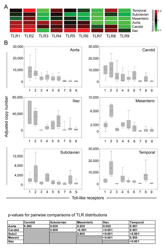
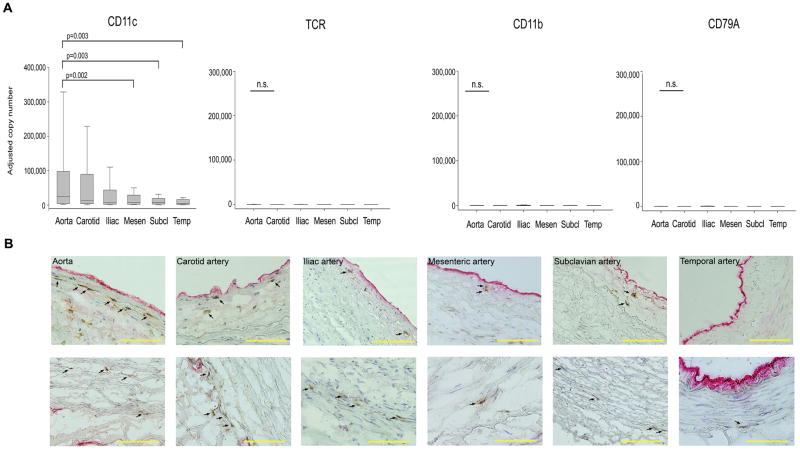
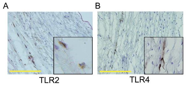
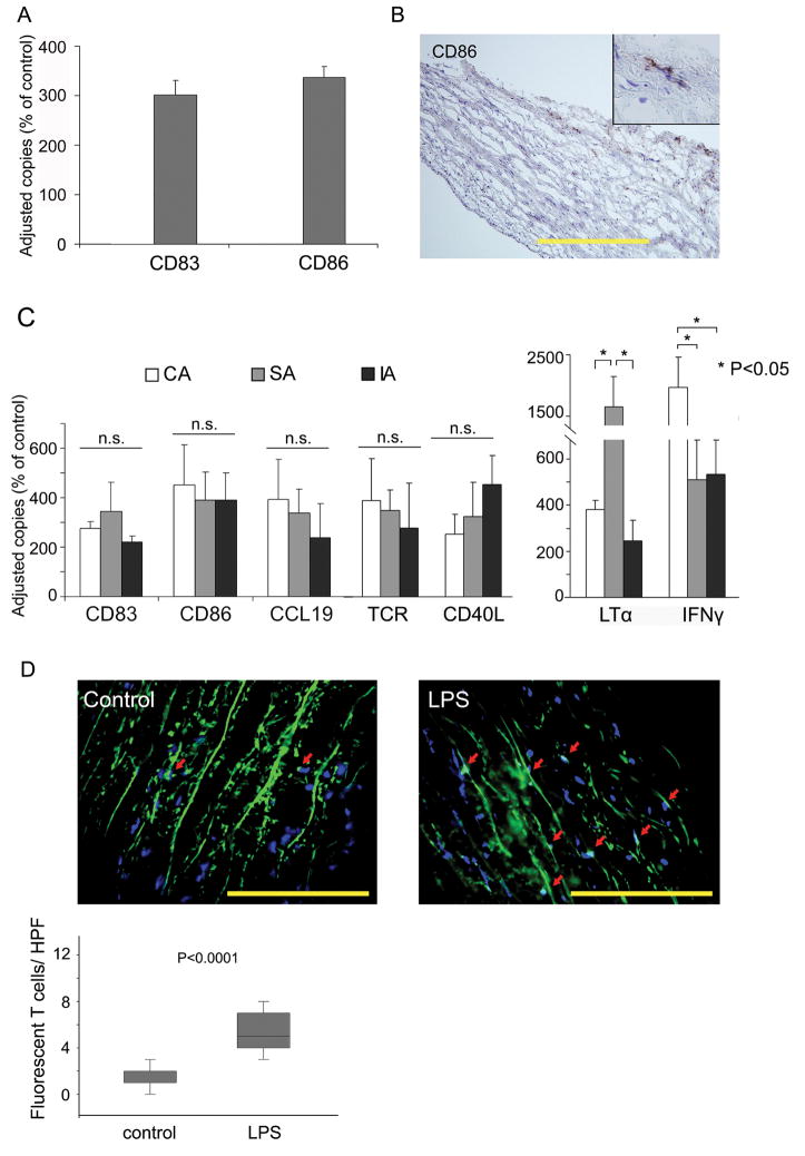
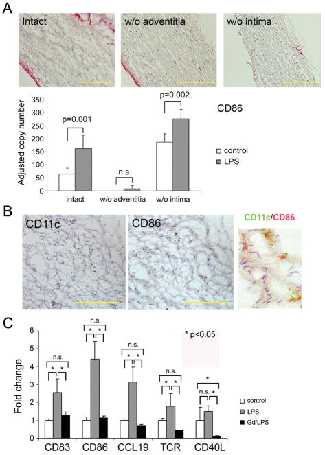
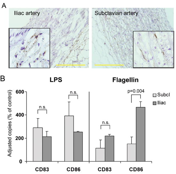
Methods and results: Gene expression analysis for pathogen-sensing Toll-like receptors (TLRs) 1 to 9 showed vessel-specific profiles, with TLR2 and TLR4 ubiquitously present, TLR7 and TLR9 infrequent, and TLR1, TLR3, TLR5, TLR6, and TLR8 expressed in selective patterns. Experiments with vessel walls stripped of the intimal or adventitial layer identified dendritic cells at the media-adventitia junction as the dominant pathogen sensors. In human artery-severe combined immunodeficiency (SCID) mouse chimeras, adoptively transferred human T cells initiated vessel wall inflammation if wall-embedded dendritic cells were conditioned with TLR ligands. Wall-infiltrating T cells displayed vessel-specific activation profiles with differential production of CD40L, lymphotoxin-alpha, and interferon-gamma. Vascular bed-specific TLR fingerprints were functionally relevant, as exemplified by differential responsiveness of iliac and subclavian vessels to TLR5 but not TLR4 ligands.

Conclusions: Populated by indigenous dendritic cells, medium and large human arteries have immune-sensing and T-cell-stimulatory functions. Each vessel in the macrovascular tree exhibits a distinct TLR profile and supports selective T-cell responses, imposing vessel-specific risk for inflammatory vasculopathies.


Figures

Figure 1
Figure 1. Expression patterns of TLR family members in distinct vascular territories
Total RNA was extracted from human aortas (n=15 different donors), carotid (n=15), iliac (n=11), mesenteric (n=15), subclavian (n=15), and temporal arteries (n=13), and transcript numbers for each TLR were measured by quantitative RT-PCR (A). Relative expression was obtained by normalizing TLR levels for each vascular bed to the median of all samples. Red fields represent higher than average expression levels; green fields denote below average transcript expression. (B) Transcript numbers for each individual TLR in each vascular bed normalized to β-actin copies are presented as box plots with medians, 25th and 75th percentiles as boxes, and 10th and 90th percentiles as whiskers. p values for pairwise comparisons of TLR expression patterns amongst the 6 arterial territories are shown.
Figure 2
Figure 2. Immune cell populations in major human arteries
(A) Samples from human arteries were collected as described in Fig. 1, and mRNA levels for the cell-type specific markers CD11c, T-cell receptor (TCR), CD11b, and the B-cell marker CD79A were quantified by RT-PCR. Results are shown as box plots with medians, 25th and 75th percentiles as boxes, and 10th and 90th percentiles as whiskers. (B) Physical localization of CD11c+ vascular dendritic cells within the arterial wall was assessed by immunohistochemistry. Frozen sections of the different vascular specimens were doublestained with antibodies specific for CD11c (brown) and the endothelial cell marker vWF (red). Vascular DC localized within the intima (top), and at the media-adventitia border (bottom) are highlighted by arrows. Scale bar: 100μm.
Figure 3
Figure 3. TLR2 and TLR4 protein expression within the normal vascular wall
Arterial cross-sections were stained with antibodies specific for TLR2 or TLR4. Representative stains for subclavian (A) and iliac artery (B) are shown. (DAB, brown, magnification ×200, inset magnifications ×400). TLR2- or TLR4-expressing cells were predominantly found along the media-adventitia border. Scale bar: 100μm.
Figure 4
Figure 4. Vascular DC sense TLR4 ligands in vitro and in vivo
(A) Subclavian arteries were with or without LPS (1μg/ml) for 12 hours in vitro. mRNA from tissue extracts were analyzed for CD83 and CD86 expression by RT- PCR. Copy numbers of the target mRNA are presented as percent of untreated control. Results from 1 of 5 experiments are shown as mean ± SD of triplicate measurements. (B) LPS-induced expression of CD86 protein was restricted to DC at the media-adventitia border. Frozen sections were stained with anti-CD86 and developed with DAB (magnification ×200, inset magnification ×400). Scale bar: 100μm. (C) Carotid (open bars), subclavian (gray bars), and iliac (black bars) arteries were engrafted into SCID mice. Chimeras received LPS (3 μg/mouse) or PBS by i.p. injection. Human CD4+ T cells were adoptively transferred 24 hours later into all mice. Grafts were explanted and markers of DC activation (CD83, CD86, CCL19) and T-cell recruitment (TCR) and in-situ activation (CD40L, LTα, IFNγ) were quantified by RT-PCR. Results are presented as percent of sham-treated control. (D) CD4 T cells were labeled with PKH67 prior to adoptive transfer. Human subclavian artery grafts were explanted, and fluorescent T cells infiltrating into the grafts were quantified in 25 randomly chosen HPF. Results are shown as box plots with medians, 25th and 75th percentiles as boxes, and 10th and 90th percentiles as whiskers. Scale bar: 50 μm.
Figure 5
Figure 5. Pathogen sensing is a function of the adventitia, not the intima
(A) Partial walls from subclavian arteries were generated by stripping off either the intima or the adventitia. Efficiency of denudation was monitored by staining for endothelial vWF (red stain). Scale bar: 100 μm. Intact and partial vessel walls were stimulated with LPS for 24 hours. CD86 mRNA was quantified by RT-PCR, and results from 5 experiments are shown as mean ± SD. (B) Induction of CD86 protein on CD11c+ adventitial DC was assessed by immunohistochemistry on serial slides. To confirm co-localization, pseudocolored sections were merged. Scale bar: 50 μm. (C) Dendritic cells were depleted from intact vessel walls by incubation with gadolinium (III) chloride (Gd). Vessels were subsequently engrafted into SCID mice. DC and T-cell functions were evaluated after LPS injection and adoptive transfer of human CD4 T cells. DC activation markers (CD83, CD86, CCL19) and T-cell markers (TCR, CD40L) were quantified in explanted human arteries by RT-PCR and are presented as fold change compared to untreated controls. Results from 1 of 3 independent experiments are shown as mean ± SD.
Figure 6
Figure 6. Vessel-specific TLR expression patterns predict responsiveness to TLR ligands
(A) Frozen sections of common iliac (left) and subclavian (right) arteries were stained with anti-TLR5 antibodies (DAB, brown). Scale bar: 100 μm. (B) Responsiveness to TLR ligands was probed by stimulating with either LPS (1 μg/mL) or flagellin (1 μg/mL) and quantified by measuring the induction of CD83 and CD86 mRNA after 24 hours stimulation in organ culture. Results are shown as percent of control.

References

    1. Wolinsky H, Glagov S. Nature of species differences in the medial distribution of aortic vasa vasorum in mammals. Circ Res. 1967;20:409–421. - PubMed
    1. Zeek PM. Periarteritis nodosa; a critical review. Am J Clin Pathol. 1952;22:777–790. - PubMed
    1. Morgan MD, Savage CO. Vasculitis in the gastrointestinal tract. Best Pract Res Clin Gastroenterol. 2005;19:215–233. - PubMed
    1. Burke AP, Tavora F, Narula N, Tomaszewski JE, Virmani R. Aortitis and ascending aortic aneurysm: description of 52 cases and proposal of a histologic classification. Hum Pathol. 2008;39:514–526. - PubMed
    1. Weyand CM, Goronzy JJ. Medium- and large-vessel vasculitis. N Engl J Med. 2003;349:160–169. - PubMed

Publication types

Substances