Review
. 2008 Sep 1;14(17):5318-24.
doi: 10.1158/1078-0432.CCR-07-5136.

Targeting the MDM2-p53 interaction for cancer therapy

Affiliations
Review

Targeting the MDM2-p53 interaction for cancer therapy

Sanjeev Shangary et al. Clin Cancer Res. .

Abstract

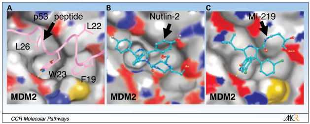
p53 is a powerful tumor suppressor and is an attractive cancer therapeutic target because it can be functionally activated to eradicate tumors. The gene encoding p53 protein is mutated or deleted in half of human cancers, which inactivates its tumor suppressor activity. In the remaining cancers with wild-type p53 status, its function is effectively inhibited through direct interaction with the human murine double minute 2 (MDM2) oncoprotein. Blocking the MDM2-p53 interaction to reactivate the p53 function is a promising cancer therapeutic strategy. This review will highlight the advances in the design and development of small-molecule inhibitors of the MDM2-p53 interaction as a cancer therapeutic approach.


Conflict of interest statement

Disclosure of Potential Conflicts of Interest

S. Wang is a consultant for Ascenta, has received a commercial research grant from Ascenta, and has an ownership interest in Ascenta.

Figures

Fig. 1
Fig. 1
Regulation of p53 and MDM2 and the outcomes of p53 activation. A, MDM2 inhibits p53 through an autoregulatory loop. MDM2 directly binds to the transactivation domain of p53 and inhibits its transcriptional activity, causes the ubiquitination and proteasomal degradation of p53, and exports p53 out of the nucleus which promotes p53 degradation and inhibits its activity. MDMX, a homologue of MDM2, also directly binds to the transactivation domain of p53 and inhibits p53 activity, but does not induce p53 degradation. ARF binds to MDM2 and sequesters MDM2 into the nucleolus, leading to the stabilization of p53. B, activation of p53 can also lead to induction of apoptosis via intrinsic (mitochondrial) and extrinsic (death receptor) apoptosis pathways. Apoptosis can be transcriptional-dependent or -independent because p53 itself can participate in mitochondrial mediated apoptosis through interaction with proapoptotic and antiapoptotic members of the Bcl-2 family. C, activation of p53 can halt cell cycle progression in the G1-S and G2-M boundaries of cell cycle through the up-regulation of the p21, Gadd45, and 14-3-3-σ proteins. Transition into the S-phase requires cyclin-dependent kinases (CDK), such as CDK2, which phosphorylates and inactivates Rb, rendering E2F free and transcriptionally active, leading to cell cycle progression. However, p53 activation induces the CDK inhibitor p21, which leads to cell cycle arrest. Furthermore, Cdc2/cyclinE activity is essential for entry into mitosis, and this activity can be inhibited by p21, Gadd45, and 14-3-3-σ, resulting in G2-M phase arrest. D, senescence is a potent tumor suppressor mechanism of p53. Telomere erosion, DNA damage, and oxidative stress or oncogenic stress can signal p53 activation, triggering senescence response via the p21-Rb-E2F signaling pathway. Oncogenic Ras can activate MAPkinase pathway that phosphorylates and activates p53 and also induces the expression of ARF, which in turn binds to and inhibits MDM2, leading to the up-regulation of p53 and the induction of senescence. E, p53 can suppress angiogenesis through the down-regulation of proangiogenic proteins and up-regulation of antiangiogenic proteins. In addition, p53 can bind to HIF-1α, a promoter of angiogenesis during hypoxia, and target it for degradation by MDM2. In a p53-independent manner, HIF-1α interacts with the p53-binding domain of MDM2, and transcriptionally up-regulates the vascular endothelial growth factor (VEGF), promoting angiogenesis. F, p53 plays a critical role in DNA damage repair. DNA damage and replication errors can activate ataxia telangiectasia mutated (ATM) and ataxia telangiectasia and Rad-related (ATR) kinases, which trigger several cellular responses, including DNA repair. ATM and ATR can phosphorylate DNA repair protein 53BP1 as well as induce the accumulation of p53 through phosphorylation directly or via CHK1and CHK2 kinases. p53 participates in DNA repair in a transactivation-dependent manner through the up-regulation of proteins such as p53R2 (p53-inducible small subunit of ribonucleotide reductase), p48 (gene product of DDB2 gene), and XPC (xeroderma pigmentosum group C protein), and in an independent manner through interaction with other DNA repair proteins, such as 53BP1.
Fig. 2
Fig. 2
Binding mode of (A) p53 peptide (PDB:1YCR) and (B) a Nutlin analogue (PDB:1RV1), and (C) predicted binding model of MI-219 to MDM2. Side chains of p53 residues involved in the MDM2-p53 interaction are shown in stick representation, whereas Nutlin-2, an analogue of Nutlin-3, and MI-219 are shown in ball-and-stick representation. Nutlin-2 is shown with carbons in cyan, nitrogen in blue, oxygen in red and bromine in brown. MI-219 is shown with carbons in cyan, nitrogen in blue, oxygen in red, fluorine in light blue, and chlorine in green. The surface representation of MDM2 in each case is shown with carbons in gray, nitrogen in blue, oxygen in red, and sulfur in yellow. Hydrogen bonds are depicted with yellow lines and hydrogen atoms are excluded for clarity. The figures were generated by Dr. Denzil Bernard using the program Pymol.

References

    1. Teodoro JG, Evans SK, Green MR. Inhibition of tumor angiogenesis by p53: a new role for the guardian of the genome. JMol Med. 2007;85:1175–86. - PubMed
    1. Fridman JS, Lowe SW. Control of apoptosis by p53. Oncogene. 2003;22:9030–40. - PubMed
    1. Vousden KH, Lu X. Live or let die: the cell’s response to p53. Nat Rev Cancer. 2002;2:594–604. - PubMed
    1. Lane DP, Crawford LV. Tantigen is bound to a host protein in SV40-transformed cells. Nature. 1979;278:261–3. - PubMed
    1. DeLeo AB, Jay G, Appella E, Dubois GC, Law LW, Old LJ. Detection of a transformation-related antigen in chemically induced sarcomas and other transformed cells of the mouse. Proc Natl Acad Sci U S A. 1979;76:2420–4. - PMC - PubMed

Publication types

MeSH terms

Substances