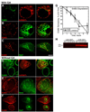
. 2008 Oct 1;121(Pt 19):3155-66.
doi: 10.1242/jcs.020404. Epub 2008 Sep 2.

Clathrin-independent endocytosis of ErbB2 in geldanamycin-treated human breast cancer cells

Affiliations

Clathrin-independent endocytosis of ErbB2 in geldanamycin-treated human breast cancer cells

Daniel J Barr et al. J Cell Sci. .

Abstract

The epidermal growth factor (EGF)-receptor family member ErbB2 is commonly overexpressed in human breast cancer cells and correlates with poor prognosis. Geldanamycin (GA) induces the ubiquitylation, intracellular accumulation and degradation of ErbB2. Whether GA stimulates ErbB2 internalization is controversial. We found that ErbB2 was internalized constitutively at a rate that was not affected by GA in SK-BR-3 breast cancer cells. Instead, GA treatment altered endosomal sorting, causing the transport of ErbB2 to lysosomes for degradation. In contrast to earlier work, we found that ErbB2 internalization occurred by a clathrin- and tyrosine-kinase-independent pathway that was not caveolar, because SK-BR-3 cells lack caveolae. Similar to cargo of the glycosylphosphatidylinositol (GPI)-anchored protein-enriched early endosomal compartment (GEEC) pathway, internalized ErbB2 colocalized with cholera toxin B subunit, GPI-anchored proteins and fluid, and was often seen in short tubules or large vesicles. However, in contrast to the GEEC pathway in other cells, internalization of ErbB2 and fluid in SK-BR-3 cells did not require Rho-family GTPase activity. Accumulation of ErbB2 in vesicles containing constitutively active Arf6-Q67L occurred only without GA treatment; Arf6-Q67L did not slow transport to lysosomes in GA-treated cells. Further characterization of this novel clathrin-, caveolae- and Rho-family-independent endocytic pathway might reveal new strategies for the downregulation of ErbB2 in breast cancer.


Figures
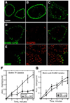
Fig. 1
Fig. 1
Effect of bound antibodies, GA, and CPZ on ErbB2 localization in SKBr3 cells. (A) ErbB2 in fixed, permeabilized cells detected by IF. (B) Cells were warmed for 2 hours after binding Fl-anti-ErbB2 before fixation. (C) Cells were treated with GA for 2 hours before detecting ErbB2 by IF. (D,E) Cells were pre-treated for 45 minutes at 37°C with GA, with (D) or without (E) 12 µg/ml CPZ, before binding anti-ErBb2 antibodies and AF-594-Tf for 1 hour on ice and warming for 2 minutes with the same drugs. Cells were acid-washed and processed for IF, detecting ErbB2 with the AF-488 Zenon mouse IgG labeling kit. (D,E) ErbB2, left; Tf, center; merged images, right. Scale bar; 10 µm. (F,G) Internalization of biotinylated Tf (F) or biotinylated anti-ErbB2 antibodies (G) was measured by CELISA after treatment with GA (circles) or both GA and CPZ (squares). Values shown are the mean +/− s.e.m. of 3 experiments.
Fig. 2
Fig. 2
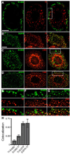
Localization of internalized ErbB2, Rh-Tf, EGFR, and clathrin. SKBr3 cells were pre-treated with GA for 1 hour before binding Fl-anti-ErbB2 (A,C,E,F) or Fl-anti-EGFR (B,D,G) and warming for 5 minutes, with Rh-Tf in A,B,E, acid washing, and IF. (C,D) Clathrin heavy chain (CHC) was detected by IF. (E–G) high-magnification views of boxed regions in A,C,D respectively. Right-hand panels in A–D and bottom panels in E–G show merged images. (A) epifluorescence images; all other panels show maximum intensity projections of deconvolved Z-stacks. Scale bars; 10 µm. Bar in B applies to panels B–D. (H) Quantitation of colocalization of Rh-Tf with ErbB2 or EGFR, in cells treated as in A,B, except that internalization was for 2 minutes.
Fig. 3
Fig. 3
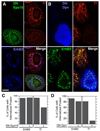
Dominant-negative forms of Eps15 and dynamin inhibit internalization of Rh-Tf but not ErbB2. SKBr3 cells transfected with EGFP-DN-Eps15 (A,C) or HA-DN-dynamin-1 (B,D) were pretreated with GA for 2 hours before binding unlabeled anti-ErbB2 antibodies (A,C) or Fl-anti-ErbB2 antibodies (B,D), warmed for 30 minutes with Rh-Tf, fixed and permeabilized. (A,B) EGFP-DN-Eps15 (A, green) or HA-DN-dynamin (B, blue) are shown with Rh-TF and ErbB2 in deconvolved images, each from a Z-stack of a field in which one cell expressed DN-Eps15 (A) or DN-dynamin (B). ErbB2 was detected with AF-350 goat anti-mouse antibodies (A,C; blue) or by fluorescein fluorescence (B,D; green). Scale bar; 10 µm. (C,D) ErbB2 and Rh-Tf internalization in cells expressing EGFP-DN-Eps15 (C) or DN-dynamin (D), and untransfected cells on the same coverslips. Cells showing at least three intracellular puncta were scored positive. Numbers shown are averages of two experiments (counting at least 100 transfected and 100 untransfected cells in each experiment) that varied by <10%.
Fig. 4
Fig. 4
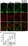
Genistein inhibits EGF-stimulated tyrosine kinase activity but not ErbB2 internalization. SKBr3 cells were serum-starved overnight, treated as described for individual panels, fixed, and permeabilized. Cells were treated with: (A) GA for 2 hours; (B) 100 ng/ml EGF for 10 minutes; (C) GA for 2 hours, with 100 ng/ml EGF added for the last 10 minutes; and (D) 100 µg/ml genistein for 1 hour, then GA added for another 2 hours, and 100 ng/ml EGF added for the last 10 minutes. Deconvolved images from Z-stacks are shown. ErbB2 (green, top) and P-Tyr (red, middle) were detected by IF. Bottom; merged images. Scale bar; 10 µm. (E) SKBr3 cells were pre-treated with GA (circles) or GA + 100 µg/ml genistein (squares) for 45 minutes before binding biotinylated anti-ErbB2 antibodies and warming for 0–5 minutes. Internalized antibodies were quantitated by CELISA. Values shown are the mean +/− s.e.m. of 3 experiments.
Fig. 5
Fig. 5
Internalized ErbB2 colocalizes with AF-594-CTxB, GPI-anchored proteins, and dextran in GA-treated SKBr3 cells. Cells were pre-treated with GA 1 hour, subjected to antibody and/or toxin binding, warmed for 5 minutes, acid-stripped, and fixed. (A) Fl-anti-ErbB2 and AF-594-CTxB (0.5 µg/ml) were bound to cells. A merged maximum intensity projection image of a deconvolved Z-stack is shown (ErbB2, green; AF-594-CTxB, red). Asterisks indicate region shown enlarged in B (ErbB2, left; AF-594-CTxB, middle; merged image, right). (C) Fl-anti-PLAP Fab fragments and Rh-anti-ErbB2 antibodies were bound to cells. A deconvolved image from a Z-stack, showing part of the edge of one cell, is shown. ErbB2, left; PLAP, middle; merged image, right. (D) Fl-anti-ErbB2 was bound to cells, which were warmed with 1 mg/ml FluoroRuby dextran. An epifluorescence image, showing part of the edge of one cell, is shown. ErbB2, left; dextran, middle; merged image, right. Scale bars; A, 10 µm; D (applies to B–D), 5 µm. (E) Colocalization of ErbB2 and CTxB, or ErbB2 and PLAP, in cells treated as in A–C (except that internalization was for 2 minutes) was quantitated. To measure colocalization of ErbB2 and Thy1.1, SKBr3 cells transfected with Thy1.1 were treated with GA 1 hour. Fl-anti-ErbB2 and AF594-anti-Thy1 Fab fragments were bound on ice, and cells warmed for 2 minutes. Residual surface-bound antibodies were acid-stripped before fixation, visualization, and quantitation.
Fig. 6
Fig. 6
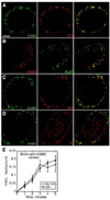
Internalized ErbB2 colocalizes with CTxB, GPI-anchored proteins, and dextran in SKBr3 cells without GA. (A–D) Cells were subjected to antibody and/or toxin binding on ice for 1 hour, warmed for 2 minutes, acid-stripped, and fixed. (A) Fl-anti-ErbB2 and AF-594-CTxB (0.5 µg/ml) were bound to cells. (B) Fl-anti-PLAP Fab fragments and Rh-anti-ErbB2 antibodies were bound. (C) Fl-anti-ErbB2 was bound to cells, which were warmed with 1 mg/ml FluoroRuby dextran. (D) Fl-anti-ErbB2 and Rh-Tf were bound. (A–D) Deconvolved images from Z-stacks are shown. ErbB2, left; CTxB, PLAP, dextran, or Tf, middle; merged images, right. Scale bar; 10 µm. (E) Internalization of biotinylated anti-ErbB2 antibodies was measured by CELISA in cells treated with (squares) or without (circles) GA. Values shown are the mean +/− s.e.m. of 3 experiments.
Fig. 7
Fig. 7
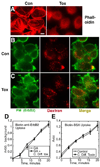
C. difficile toxin B does not inhibit internalization of ErbB2 or fluid in SKBr3 cells. (A) SKBr3 cells grown for 48 hours on poly-Lys-coated coverslips were left untreated (Con) or treated 2 hours with 0.5 µg/ml C. difficile toxin B (Tox), fixed, permeabilized and incubated with rhodamine phalloidin (4 U/ml). Stress fibers were seen in about half of the control cells and <1% of treated cells. (B,C) Serum-starved SKBr3 cells on poly-Lys-coated coverslips were left untreated (B) or treated 2 hours with 0.5 µg/ml C. difficile toxin B (C) before addition of FluoroRuby dextran (1 mg/ml) for 10 minutes. After fixation, surface morphology was visualized with anti-ErbB2 antibodies and green secondary antibodies. Scale bar; 10 µm. (D,E) Internalization of biotinylated anti-ErbB2 antibodies (D) or biotinylated BSA (E) was measured by CELISA in serum-starved SKBr3 cells treated with GA (D, circles), GA + C. difficile toxin B (D, squares), C. difficile toxin alone (E, squares), or left untreated (E, circles) as described in Methods. Where appropriate, cells were pre-incubated with C. difficile toxin B (0.5 µg/ml) for 2 hours at 37°C with addition of GA for the last 45 minutes. Values shown are the mean +/− s.e.m. of 3 experiments.
Fig. 8
Fig. 8
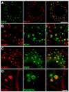
ErbB2 is delivered to early endosomes after GA treatment. SKBr3 cells were left untransfected (A,B) or transiently transfected with GFP-Rab5 (C) or GFP-Rab5Q79L (D), and then treated with GA for 2 hours (with Rh-Tf added for the last 30 minutes in A), fixed, and permeabilized. (A–D) Left panels; ErbB2, detected with polyclonal antibodies. Center panels: (A) Rh-Tf fluorescence; (B) endogenous EEA1; (C,D) GFP fluorescence. Right panels; merged images. (A) deconvolved image from a Z stack; (B–D) epifluorescence images. Scale bars; A, 10 µm; D (applies to B–D), 5 µm.
Fig. 9
Fig. 9
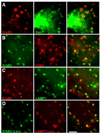
ErbB2 is delivered to late endosomes and lysosomes after GA treatment. After transient expression of GFP-Rab7 (A only), SKBr3 cells were treated with GA for 5 hours (together with 0.1 mg/ml leupeptin, D only), fixed, and permeabilized. Left panels; ErbB2 was detected with polyclonal (A,B) or monoclonal (C,D) antibodies and appropriate secondary antibodies. Middle panels: (A) Rab7 (GFP fluorescence); (B) endogenous CD63; (C,D) endogenous LAMP1. Right panels; merged images. Epifluorescence images are shown. Scale bar; 5 µm.
Fig. 10
Fig. 10
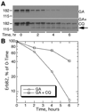
GA-induced ErbB2 degradation is sensitive to chloroquine. SKBr3 cells were incubated with GA with or without CQ for the times indicated and lysed. Proteins were separated by SDS-PAGE and transferred to membranes for Western blotting and detection of ErbB2. (A) Western blots. Top, GA alone; bottom, GA and chloroquine (CQ). Arrow; ca. 135 kDa ErbB2 fragment. (B) Bands were quantitated by scanning densitometry and plotted as % of 0-time signal remaining at each time.
Fig. 11
Fig. 11
ErbB2 accumulates in Arf-Q67L-positive endosomes only without GA. (A–F) SKBr3 cells were transfected with Arf6-Q67L, alone (A,D) or with GFP-Rab5 (B,E) or GFP-Rab7 (C,F). Fl-anti-ErbB2 (A,D) or unlabeled anti-ErbB2 antibodies (B,C,E,F) were bound for 1 hour before cells were warmed for 2 hours with (A–C) or without (D–F) GA. Internalized anti-ErbB2 was detected in fixed and permeabilized cells by Fl-anti-ErbB2 fluorescence (A,D) or with Texas red goat-anti-mouse antibodies (B,C,E,F). Although Arf6Q67L was not visualized in B,C,E, or F, vacuoles characteristic of Arf6-Q67L expression were seen. Scale bar; 10 µm. (G,H) COS-7 cells were transfected with ErbB2 alone or together with Arf6-Q67L as indicated. Cells were incubated with GA for the indicated times and solubilized in gel loading buffer. Proteins were separated by SDS-PAGE and transferred to nitrocellulose. ErbB2 and Arf6-Q67L were detected by immunoblotting as described in Methods. (H) A representative Arf6-Q67L blot is shown, demonstrating expression in co-transfected cells (+Arf6-Q67L), but not in cells expressing ErbB2 alone (−Arf6-Q67L).

References

    1. Arreaza G, Melkonian KA, LaFevre-Bernt M, Brown DA. Triton X-100-resistant membrane complexes from cultured kidney epithelial cells contain the Src-family protein tyrosine kinase p62yes. J. Biol. Chem. 1994;269:19123–19127. - PubMed
    1. Austin CD, De Mazière AM, Pisacane PI, van Dijk SM, Eigenbrot C, Sliwkowski MX, Klumperman J, Scheller RH. Endocytosis and sorting of ErbB2 and the site of action of cancer therapeutics trastuzumab and geldanamycin. Mol. Biol. Cell. 2004;15:5268–5282. - PMC - PubMed
    1. Austin CD, Wen X, Gazzard L, Nelson C, Scheller RH, Scales SJ. Oxidizing potential of endosomes and lysosomes limits intracellular cleavage of disulfide-based antibody–drug conjugates. Proc. Natl. Acad. Sci. USA. 2005;102:17987–17992. - PMC - PubMed
    1. Baulida J, Kraus MH, Alimandi M, DiFiore PP, Carpenter G. All ErbB receptors other than the epidermal growth factor receptor are endocytosis impaired. J. Biol. Chem. 1996;271:5251–5257. - PubMed
    1. Beerli RR, Graus-Porta D, Woods-Cook K, Chen X, Yarden Y, Hynes NE. Neu differentiation factor activation of ErbB-3 and ErbB-4 is cell specific and displays a differential requirement for ErbB-2. Mol. Cell. Biol. 1995;15:6496–6505. - PMC - PubMed

Publication types

MeSH terms