

MRI correlates of neurofibrillary tangle pathology at autopsy: a voxel-based morphometry study
- PMID: 18765650
- PMCID: PMC2676950
- DOI: 10.1212/01.wnl.0000324924.91351.7d
MRI correlates of neurofibrillary tangle pathology at autopsy: a voxel-based morphometry study
Abstract
Background: Neurofibrillary tangles (NFTs), composed of hyperphosphorylated tau proteins, are one of the pathologic hallmarks of Alzheimer disease (AD). We aimed to determine whether patterns of gray matter atrophy from antemortem MRI correlate with Braak staging of NFT pathology.
Methods: Eighty-three subjects with Braak stage III through VI, a pathologic diagnosis of low- to high-probability AD, and MRI within 4 years of death were identified. Voxel-based morphometry assessed gray matter atrophy in each Braak stage compared with 20 pathologic control subjects (Braak stages 0 through II).
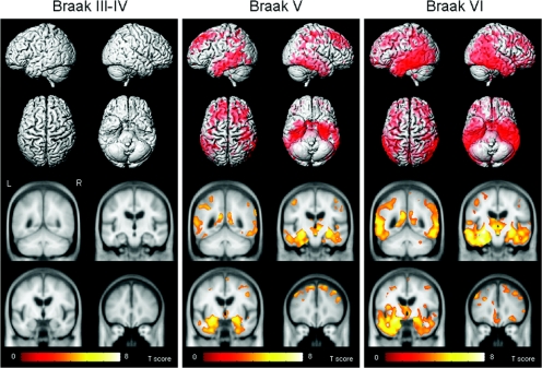
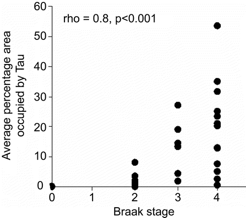
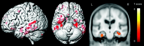
Results: In pairwise comparisons with Braak stages 0 through II, a graded response was observed across Braak stages V and VI, with more severe and widespread loss identified at Braak stage VI. No regions of loss were identified in Braak stage III or IV compared with Braak stages 0 through II. The lack of findings in Braak stages III and IV could be because Braak stage is based on the presence of any NFT pathology regardless of severity. Actual NFT burden may vary by Braak stage. Therefore, tau burden was assessed in subjects with Braak stages 0 through IV. Those with high tau burden showed greater gray matter loss in medial and lateral temporal lobes than those with low tau burden.
Conclusions: Patterns of gray matter loss are associated with neurofibrillary tangle (NFT) pathology, specifically with NFT burden at Braak stages III and IV and with Braak stage itself at higher stages. This validates three-dimensional patterns of atrophy on MRI as an approximate in vivo surrogate indicator of the full brain topographic representation of the neurodegenerative aspect of Alzheimer disease pathology.
Figures
References
-
- Braak H Braak E Neuropathological stageing of Alzheimer-related changes. Acta Neuropathol (Berl) 1991;82:239–259. - PubMed
-
- Fox NC Warrington EK Freeborough PA et al. Presymptomatic hippocampal atrophy in Alzheimer’s disease: a longitudinal MRI study. Brain 1996. 119pt 62001 2007 - PubMed
-
- Fox NC Crum WR Scahill RI et al. Imaging of onset and progression of Alzheimer’s disease with voxel-compression mapping of serial magnetic resonance images. Lancet. 2001;358:201–205. - PubMed
-
- Matsuda H Kitayama N Ohnishi T et al. Longitudinal evaluation of both morphologic and functional changes in the same individuals with Alzheimer’s disease. J Nucl Med. 2002;43:304–311. - PubMed
Publication types
MeSH terms
Substances
Grants and funding
LinkOut - more resources
Full Text Sources
Other Literature Sources
Medical