. 2008 Oct 31;283(44):30363-75.
doi: 10.1074/jbc.M804392200. Epub 2008 Sep 2.

Up-regulating sphingosine 1-phosphate receptor-2 signaling impairs chemotactic, wound-healing, and morphogenetic responses in senescent endothelial cells

Affiliations

Up-regulating sphingosine 1-phosphate receptor-2 signaling impairs chemotactic, wound-healing, and morphogenetic responses in senescent endothelial cells

Rosendo Estrada et al. J Biol Chem. .

Abstract

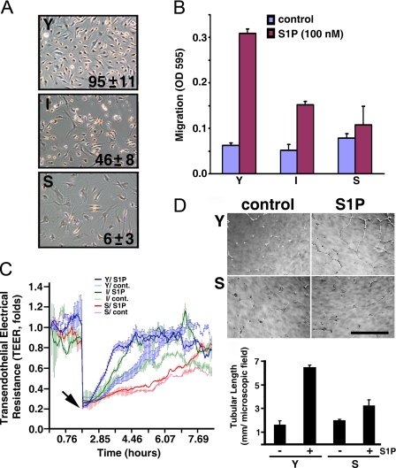
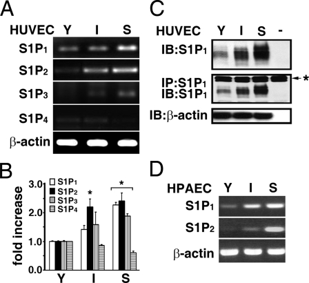
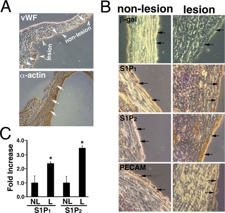
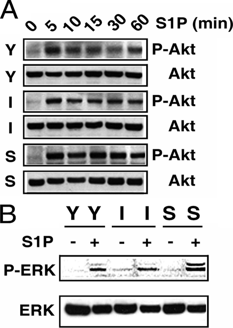
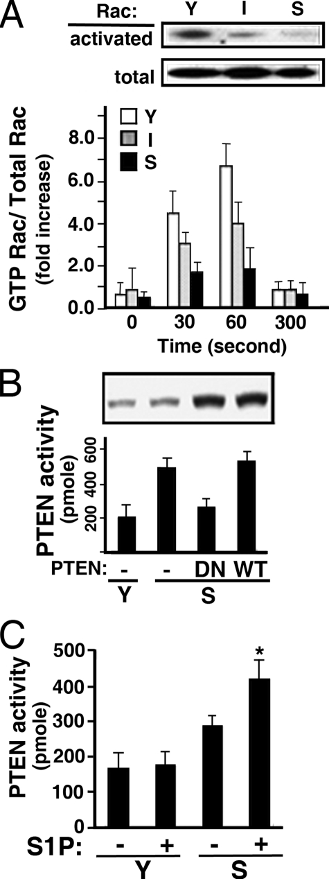
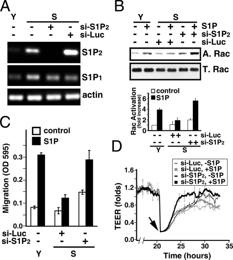
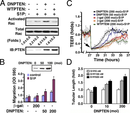
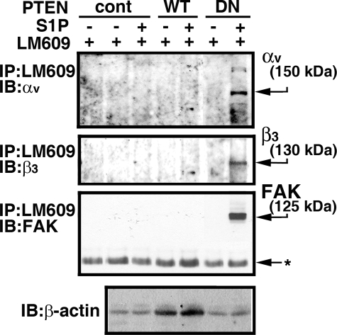
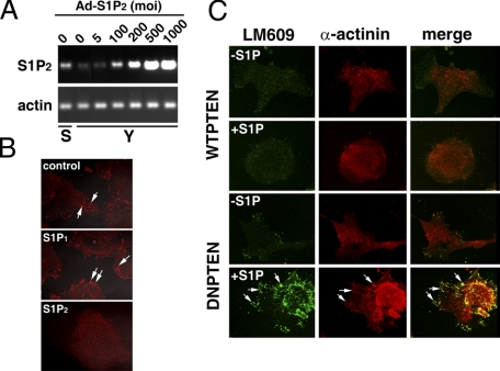
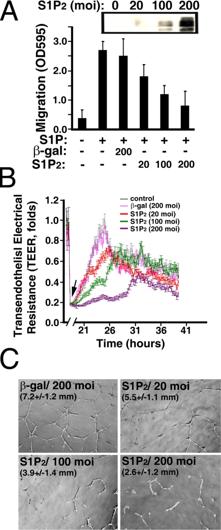
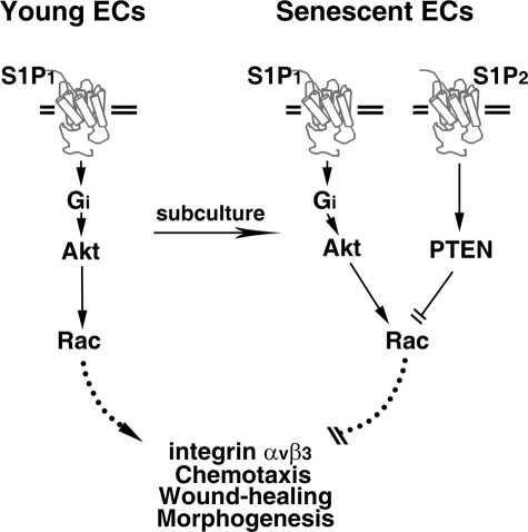
Vascular endothelial cells (ECs) have a finite lifespan when cultured in vitro and eventually enter an irreversible growth arrest state called "cellular senescence." It has been shown that sphingolipids may be involved in senescence; however, the molecular links involved are poorly understood. In this study, we investigated the signaling and functions of sphingosine 1-phosphate (S1P), a serum-borne bioactive sphingolipid, in ECs of different in vitro ages. We observed that S1P-regulated responses are significantly inhibited and the S1P(1-3) receptor subtypes are markedly increased in senescent ECs. Increased expression of S1P(1) and S1P(2) was also observed in the lesion regions of atherosclerotic endothelium, where senescent ECs have been identified in vivo. S1P-induced Akt and ERK1/2 activation were comparable between ECs of different in vitro ages; however, PTEN (phosphatase and tensin homolog deleted on chromosome 10) activity was significantly elevated and Rac activation was inhibited in senescent ECs. Rac activation and senescent-associated impairments were restored in senescent ECs by the expression of dominant-negative PTEN and by knocking down S1P(2) receptors. Furthermore, the senescent-associated impairments were induced in young ECs by the expression of S1P(2) to a level similar to that of in vitro senescence. These results indicate that the impairment of function in senescent ECs in culture is mediated by an increase in S1P signaling through S1P(2)-mediated activation of the lipid phosphatase PTEN.


Figures

FIGURE 1.
FIGURE 1.
S1P-mediated endothelial functions are impaired in senescent ECs. A, proliferative capabilities of young (Y, CPDL = 15), intermediate (I, CPDL = 35), and senescent (S, CPDL = 60) ECs. Cells were labeled with BrdUrd reagent for 24 h. Subsequently, cells were immunostained with anti-BrdUrd following the manufacturer's instructions (BrdUrd staining kit, Zymed Laboratories Inc.). Mean ± S.E. represents percentage of BrdUrd incorporated nuclei from 3-5 microscopic fields of duplicate experiments. B, chemotactic responses were measured by a Transwell migration assay as described under “Experimental Procedures.” Note that S1P (100 nm)-induced chemotactic responses were inversely related to in vitro ages of cultured ECs. C, young, intermediate, and senescent ECs were plated into each well of 8W1E arrays and incubated at 37 °C to allow the transendothelial electrical resistance (TEER) to reach equilibrium. 24 h later, the cells attached to the microelectrodes were killed by applying 5 V of electrical current for 30 s (arrow), which resulted in an abrupt impedance drop. Subsequently, endothelial wound-healing responses in the presence or absence of 100 nm S1P were determined in real-time by measuring the recovery of TEER, an indicator of endothelial migration into the wounded area (29). D, morphogenesis of young and senescent ECs on matrigel (upper panel) and quantitative analysis of tubular length (lower panel) in the presence or absence of S1P (200 nm) were performed as described (12, 13). Scale bar, 318 μm.
FIGURE 2.
FIGURE 2.
Expression of S1P family receptor subtypes in ECs with different in vitro ages. The expression of S1P receptor subtypes in young, intermediate, and senescent HUVECs was measured by RT-PCR (A) and real-time qPCR (B) as described under “Experimental Procedures.” S1P1 expression was age-dependently increased in cultured HUVECs, and S1P2 expression was barely detected in young ECs, whereas it was markedly increased in intermediate and senescent ECs. Similarly, S1P3 was barely detected in young ECs and significantly up-regulated in senescent ECs. A low level of S1P4 receptor was detected in young and intermediate ECs and is absent in senescent ECs. S1P5 was not detected in ECs with different in vitro ages. The data are from a representative experiment, which was repeated six times with identical results. *, p < 0.01 (t test). C, endothelial extracts were immunoblotted (upper panel), or immunoprecipitated followed by Western-blotting with anti-S1P1 (middle panel). Immunoblotting with anti-β-actin was used as an internal control for loading (lower panel). The result is representative of triplicate experiments. -, assays were performed without endothelial extracts; *, immunoglobulin heavy chain used for immunoprecipitation assays. D, S1P1 and S1P2 expression in HPAECs with different in vitro ages. S1P1 expression was age-dependently increased in HPAECs. Also, S1P2 expression was significantly induced in senescent HPAECs and was undetected in young HPAECs.
FIGURE 3.
FIGURE 3.
S1P2 receptors are up-regulated in the endothelium at the atherosclerotic lesion. The aorta segments were prepared from atherosclerotic mice (∼25 weeks old) as described previously (34). The atherosclerotic lesion and non-lesion regions (arrowheads, top, panel A) of the aorta were identified as reported previously (34). The endothelium was identified by staining with anti-Von Willebrand factor (vWF, arrows, top, panel A) and anti-PECAM (arrows, bottom row, panel B). The smooth muscle layers were identified by anti-α-actin staining (arrows, bottom, panel A). B, the non-lesion and lesion regions of atherosclerotic aorta were examined for expression of SA-β-galactosidase activity (top row), S1P1 (second row), and S1P2 (third row). SA-β-galactosidase activity was observed in the endothelium of lesion regions (blue color indicated by arrows) and absent in the non-lesion regions. A small increase of S1P1 expression was observed in lesion regions compared with the non-lesion regions (brown color indicated by arrows, second row). The expression of S1P2 receptors was not detected in the endothelium of the non-lesion regions of the aorta (light brown indicated by arrows, left panel, third row). In contrast, S1P2 receptors were markedly up-regulated in the endothelium of atherosclerotic lesions (dark brown indicated by arrows, right panel, third row). C, quantitation of S1P1 and S1P2 expression. The expression of S1P1 and S1P2 receptors in lesion and non-lesion regions of the atherosclerotic endothelium was quantitated by MetaMorph software (MetaMorph Version 6.1, Molecular Devices) (35). Data represent mean ± S.E. of six microscopic fields. Both S1P1 and S1P2 receptors are significantly up-regulated in the lesion regions of atherosclerotic endothelium (*, p < 0.05, t test). NL, non-lesion; L, lesion regions.
FIGURE 4.
FIGURE 4.
Akt and ERK1/2 are effectively activated in senescent ECs. Young (Y), intermediate (I), and senescent (S) ECs (8 × 10⁵ cells in 100-mm dish) were plated for 3 days. Cells were then serum-starved in plain M199 medium for 2 h followed by stimulation with S1P (200 nm) for various times. Extracts were then probed with phospho-specific Akt (panel A) or phosphospecific ERK (panel B) antibody. Subsequently, nitrocellulose membranes were stripped and re-probed with anti-Akt (panel A) or anti-ERK (panel B) to detect the quantities of endogenous Akt or ERK polypeptides. The experiment was repeated three times with identical results.
FIGURE 5.
FIGURE 5.
Elevated PTEN and diminished Rac activities in senescent ECs. A: Upper panel, cellular lysates were prepared from normally growing young (Y), intermediate (I), and senescent (S) ECs. The activated Rac polypeptides were isolated by glutathione S-transferase-p21-activated kinase-bound Sepharose beads as described under “Experimental Procedures” (upper blot). Western blots of the cellular extracts (50 μg) with anti-Rac shows that the quantities of endogenous Rac polypeptides were not changed in ECs with different in vitro ages (lower blot). Lower panel, young, intermediate, and senescent ECs were plated for 3 days followed by serum starvation in plain M199. After stimulating with S1P (100 nm) for the indicated times, Rac activation was measured as described above. The immunoblot intensity was quantitated with a Typhoon densitometer equipped with ImageQuant software. The histogram shows S1P-stimulated Rac activation represented as the ratio of activated Rac/total cellular Rac (mean ± S.E., n = 3). B, cellular extracts of normal growing young and senescent ECs were immunoprecipitated with anti-PTEN. The phosphatase activity of precipitated PTEN was then measured as described under “Experimental Procedures.” Alternatively, senescent ECs were transduced with adenoviral particles carrying dominant-negative or wild-type PTEN (200 m.o.i.) for 16 h. The PTEN activity was then measured. Inset, Western blot shows the endogenous or transduced PTEN polypeptides. The data are the mean ± S.E. of triplicate determinants. C, young and senescent ECs were plated for 3 days followed by serum starvation in plain M199. After stimulating with S1P (100 nm, 15 min), PTEN activity was measured as described above. The data represents the mean ± S.E. of triplicate determinants (*, t test; p < 0.01).
FIGURE 6.
FIGURE 6.
S1P2 knockdown restores the impaired activities of senescent ECs. A, senescent ECs were transduced with lentiviral particles carrying siRNA oligonucleotides for Luciferase or S1P2 receptor. RT-PCR measurement shows the efficacy of S1P2 knockdown. Note that the expression of S1P2 in senescent ECs transduced with a si-S1P2 construct was completely inhibited, whereas an irrelevant siRNA (si-Luc) had no effect on S1P2 knockdown. Also note that transduction of si-S1P2 has no nonspecific off-target effects on the expression of S1P1 and actin. B, senescent ECs were transduced with lentiviral particles carrying si-S1P2 or si-Luc. Cells were grown for 3 days and serum-starved in plain M199 for 2 h. Subsequently, cells were stimulated with 200 nm S1P for 1 min, and Rac activity was measured as described above. Fold increase, mean ± S.E. of three determinants. C, endothelial migration was measured in young, si-S1P2-transduced, or si-Luc-transduced senescent ECs in the presence or absence of S1P (100 nm) stimulation. The data are mean ± S.E. of three determinants. D, si-S1P2- or si-Luc-transduced senescent ECs were plated in the ECIS array. S1P-mediated wound-healing response was measured as described above. Arrow, S1P (200 nm) was added immediately after the endothelial monolayers were injured by elevated electrical voltage. Data are the mean ± S.E. of triplicate determinants of a representative experiment, which was repeated three times with identical results.
FIGURE 7.
FIGURE 7.
The impaired activities of senescent ECs were restored by dominant negative PTEN. A, senescent ECs (CPDL = 60) were transduced with adenoviral particles (200 m.o.i.) carrying wild-type (WTPTEN) or dominant negative PTEN (DNPTEN) vectors for 16 h. After S1P stimulation (200 nm, 1 min), Rac activation was measured as described above. Folds, mean ± S.E. of three determinants. Lower panel, Western blot of cellular extracts shows the expression of endogenous or transduced PTEN polypeptides. B, senescent ECs were transduced with adenoviral particles carrying β-galactosidase or DNPTEN vectors for 16 h. Subsequently, the chemotactic responses were measured by Transwell migration assay in the presence or absence of S1P (200 nm). Inset, Western blot of cellular extracts shows the expression of transduced dominant negative PTEN polypeptides. The data shows the mean ± S.E. of three determinants. C, senescent ECs were grown to confluence in the ECIS array. Cells were then transduced with adenoviral particles carrying β-galactosidase, WTPTEN, and DNPTEN vectors for 16 h. After creating the wound by elevating voltage (arrow), S1P-mediated wound-healing responses were monitored in real-time by the increase of TEER. D, senescent ECs were infected with adenoviral particles carrying β-galactosidase or DNPTEN vectors. Cells were plated onto three-dimensional Matrigel for 24 h in the presence or absence of the indicated concentrations of S1P. The tubular lengths were quantitated and shown as mean ± S.E. of 3-5 microscopic fields of a representative experiment, which was repeated three times with similar results.
FIGURE 8.
FIGURE 8.
Transduction of dominant-negative PTEN restores S1P-stimulated integrin αvβ3 activation in senescent ECs. Senescent ECs were transduced with adenoviral particles carrying empty vector (cont), wild-type (WT), or dominant-negative (DN) PTEN constructs (200 m.o.i each) for 16 h. Cells were washed and serum starved for 2 h. Subsequently, cells were stimulated with or without S1P (500 nm) for 10 min. Cellular extracts (500 μg) were precipitated with LM609 antibody followed by immunoblotting with FAK, αv, and β3 antibodies. The migratory positions of endogenous polypeptides are indicated by arrows. Extracts (10 μg) were immunoblotted with β-actin antibody in a parallel assay to show that an equal quantity of extracts was used for the immunoprecipitation assay. Left lane, TBST/OG buffer was incubated with LM609 antibody. Note that S1P treatment was unable to induce the association of FAK with integrin αvβ3 as well as the ligation of αv and β3 subunits in the control vector and wild-type PTEN-transduced senescent ECs. In contrast, S1P stimulated the association of FAK with integrinαvβ3 as well as the ligation of αv and β3 subunits in dominant-negative PTEN-transduced senescent ECs. The data shown are from a representative experiment, which was repeated two times with similar results.
FIGURE 9.
FIGURE 9.
Ectopical expression of S1P2 inhibits integrinαvβ3 activation in young ECs. A, young ECs were transduced with increased concentrations of adenoviral particles carrying S1P2 receptors. The expression of S1P2 receptors in senescent (S) or young (Y) ECs was determined by RT-PCR. Note that the expression of S1P2 receptors in senescent ECs was attained in young ECs by transduction with ∼100 m.o.i. of adenoviral particles. B, young ECs were transduced with adenoviral particles (100 m.o.i.) carrying the β-galactosidase (top panel), S1P1 (middle panel), or S1P2 vectors (bottom panel). Cells were serum-starved, stimulated with S1P (200 nm, 37 °C for 10 min), and immunostained with mouse monoclonal LM609 (Chemicon). Note that S1P induced the activation of integrin αvβ3 at the focal contact sites of the migratory fronts in young ECs transduced with β-galactosidase and S1P1 (arrows, top two panels). In contrast, S1P was unable to activate integrin αvβ3 in young ECs transduced with adenoviral particles carrying S1P2 receptor. C, young ECs were co-transduced with adenoviral particles (100 m.o.i. each) carrying S1P2 and wild-type (first and second rows), or S1P2 and dominant-negative PTEN (third and forth rows) for 16 h. After stimulation with (second and fourth rows) or without (first and third rows) S1P (200 nm, 37 °C for 10 min), cells were immunostained with mouse monoclonal anti-LM609 (left panels) and rabbit polyclonal anti-α-actinin (middle panels). The merged images are shown in the right panels. Note that transduction of dominant-negative PTEN induced the co-localization of α-actinin and integrin αvβ3 in young ECs expressing S1P2 receptors (arrows, bottom row). In contrast, S1P treatment was unable to induce integrin αvβ3 activation and α-actinin-αvβ3 association in young ECs co-transduced with adenoviral particles carrying S1P2 and wild-type PTEN vectors.
FIGURE 10.
FIGURE 10.
S1P2 expression impairs migratory, wound-healing, and morphogenetic responses in young ECs. A, young ECs (CPDL = 15) were transduced with adenoviral particles carryingβ-galactosidase or S1P2 vectors for 16 h. Subsequently, chemotactic responses were measured by a Transwell migration assay in the presence or absence of S1P (200 nm). Inset, Western blot of cellular extracts shows the expression of transduced S1P2 polypeptides. B, young ECs were grown to confluence in the ECIS array. Subsequently, cells were transduced with adenoviral particles carrying β-galactosidase or S1P2 vectors for 16 h. After creating the wound by elevating voltage (arrow), S1P-mediated wound-healing responses were monitored in real-time by the increase of TEER. C, young ECs were infected with adenoviral particles carrying β-galactosidase or S1P2 vectors. Cells were plated onto growth factor-reduced Matrigel for 24 h in the presence of S1P (200 nm). The tubular lengths were quantitated as described (12, 13). Magnification, ×5. The data in A, B, and C are mean ± S.E. of three determinants of a representative experiment, which was repeated at least three times with similar results.
FIGURE 11.
FIGURE 11.
Model for S1P2 up-regulation impairs the activation of integrin αvβ3, chemotactic, wound-healing, and morphogenetic responses in senescent ECs. S1P1-mediated Akt/Rac signaling stimulates integrin αvβ3 activation, chemotactic, wound-healing, and morphogenetic responses in young ECs. In contrast, both S1P1 and S1P2 subtypes of S1P family receptors are up-regulated in senescent ECs. The increased expression of S1P2 antagonizes the “stimulatory” signaling mediated by S1P/S1P1 pathway via elevating cellular PTEN phosphatase activity. The S1P2-mediated PTEN activation results in the inactivation of Rac GTPase, which ultimately inhibits the integrin αvβ3 activation, migratory, wound-healing, and morphogenetic capabilities in senescent ECs.

References

    1. Moolenaar, W. H. (1999) Exp. Cell Res. 253 230-238 - PubMed
    1. Spiegel, S. (1999) J. Leukoc. Biol. 65 341-344 - PubMed
    1. Igarashi, Y., and Yatomi, Y. (1998) Acta Biochim. Pol. 45 299-309 - PubMed
    1. Hla, T., Lee, M. J., Ancellin, N., Liu, C. H., Thangada, S., Thompson, B. D., and Kluk, M. (1999) Biochem. Pharmacol. 58 201-207 - PubMed
    1. An, S., Goetzl, E. J., and Lee, H. (1998) J. Cell. Biochem. Suppl. 30-31 147-157 - PubMed

Publication types