. 2008 Nov 7;283(45):31256-67.
doi: 10.1074/jbc.M803512200. Epub 2008 Sep 2.

Drosophila MFAP1 is required for pre-mRNA processing and G2/M progression

Affiliations

Drosophila MFAP1 is required for pre-mRNA processing and G2/M progression

Ditte S Andersen et al. J Biol Chem. .

Abstract

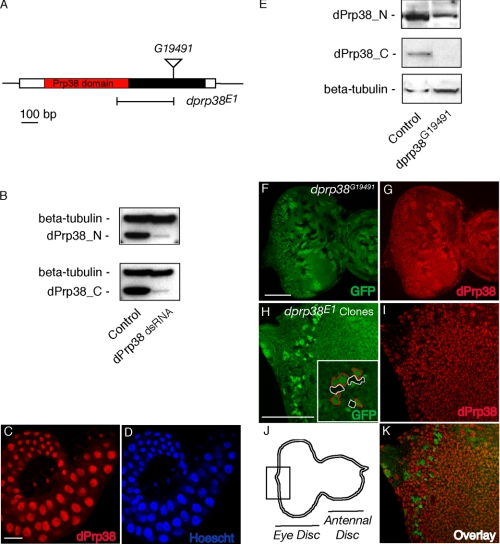
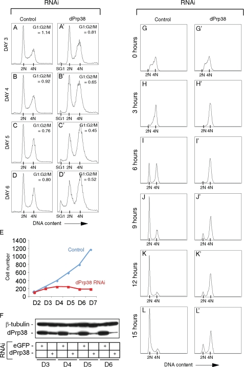
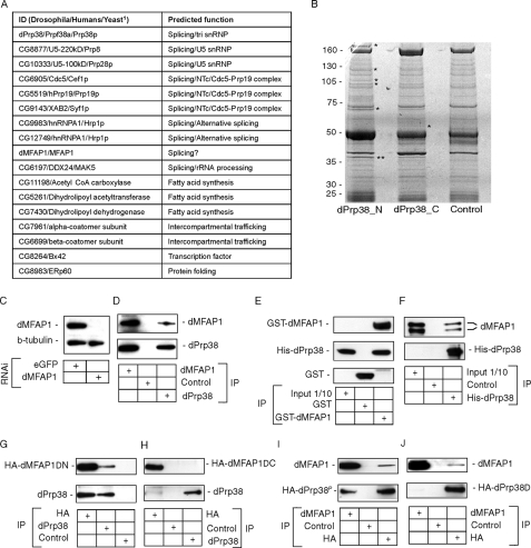
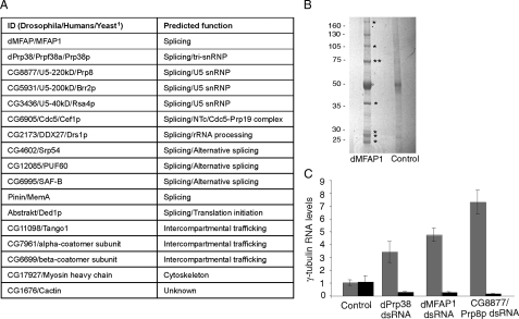
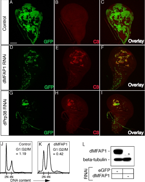
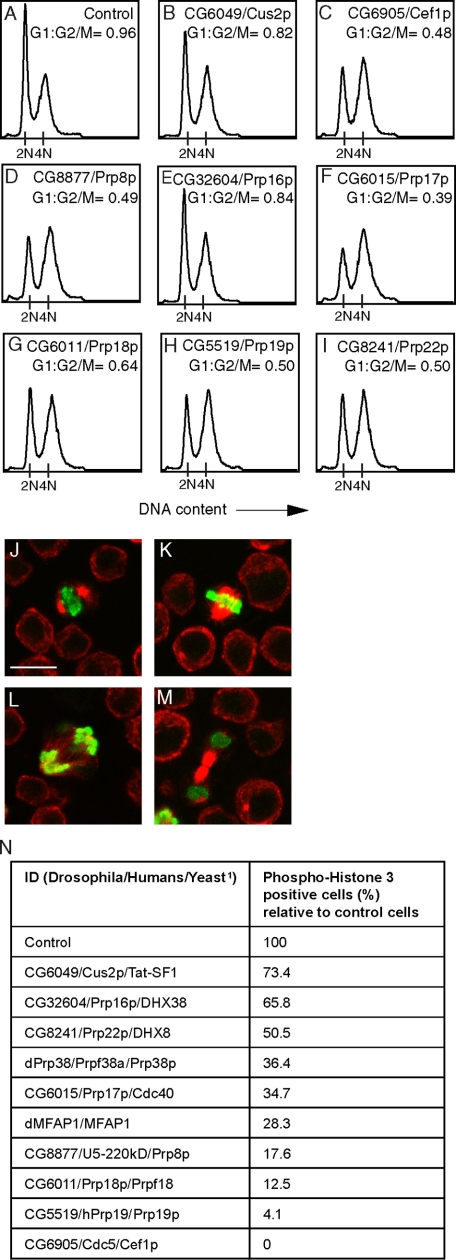
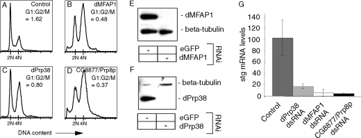
The mammalian spliceosome has mainly been studied using proteomics. The isolation and comparison of different splicing intermediates has revealed the dynamic association of more than 200 splicing factors with the spliceosome, relatively few of which have been studied in detail. Here, we report the characterization of the Drosophila homologue of microfibril-associated protein 1 (dMFAP1), a previously uncharacterized protein found in some human spliceosomal fractions ( Jurica, M. S., and Moore, M. J. (2003) Mol. Cell 12, 5-14 ). We show that dMFAP1 binds directly to the Drosophila homologue of Prp38p (dPrp38), a tri-small nuclear ribonucleoprotein component ( Xie, J., Beickman, K., Otte, E., and Rymond, B. C. (1998) EMBO J. 17, 2938-2946 ), and is required for pre-mRNA processing. dMFAP1, like dPrp38, is essential for viability, and our in vivo data show that cells with reduced levels of dMFAP1 or dPrp38 proliferate more slowly than normal cells and undergo apoptosis. Consistent with this, double-stranded RNA-mediated depletion of dPrp38 or dMFAP1 causes cells to arrest in G(2)/M, and this is paralleled by a reduction in mRNA levels of the mitotic phosphatase string/cdc25. Interestingly double-stranded RNA-mediated depletion of a wide range of core splicing factors elicits a similar phenotype, suggesting that the observed G(2)/M arrest might be a general consequence of interfering with spliceosome function.


Figures

FIGURE 1.
FIGURE 1.
dPrp38 is a nuclear protein required for developmental growth and proliferation. A, schematic of the dprp38 locus. dPrp38 is encoded by a single exon. The N-terminal part encodes a region with homology to Prp38 (red). The position of the P-element inserted in dprp38G19491 and the imprecise excision deletion mutant (dprp38E1) are indicated. B, immunoblots on cell extracts prepared from S2 cells treated with dsRNA corresponding to GFP (control) or dPrp38 probed with antibodies recognizing the C- (dPrp38_C) and N-terminal (dPrp38_N) part of dPrp38. C and D, dPrp38 is localized in the nucleus. Wild-type salivary gland tissues from third instar larvae stained with the anti-dPrp38_C antibody in red (C) or a nuclear dye (D). E, whole extracts from control adult flies or flies homozygous mutant for dprp38G19491 were used for immunoblotting with dPrp38_N (top panel), dPrp38_C (middle panel), and anti-β-tubulin (bottom panel) antibodies. F-K, eye imaginal discs from third instar larvae. Posterior is to the left. F and H, the scale bar is at 100 μm. Mitotic clones of dprp38G19491 (F and G) or dprp38E1 (H-K) mutant tissue marked by the absence of GFP (F) or two copies of GFP (H and K) and stained with the dPrp38_C antibody in red (G, I, and K). Inset in H is a blow-up of a region of the image, with dprp38 mutant areas circled in red and homozygous Minute clones in white. J, a schematic representation of the eye-antennal disc with the area in H, I, and K boxed.
FIGURE 2.
FIGURE 2.
dPrp38 is required for proliferation and G2/M progression. A-D′, cells depleted of dPrp38 gradually accumulate in G2/M and eventually become apoptotic. S2 cells were treated with dsRNA targeting eGFP (control) or dprp38 for the indicated number of days. Cells were fixed, stained with propidium iodide, and analyzed by flow cytometry. The ratio of cells in the G2/M relative to G1 phase is indicated for each treatment (G1:G2/M). The peaks corresponding to the sub-G1 (SG1), G1 (2N), and G2/M (4N) populations are indicated on the x axis. E and F, cells were counted and dPrp38 protein levels measured in parallel. E, control treated (blue) or dPrp38-depleted (red) cells were counted once a day on days 2-7 (D2-D7). F, immunoblotting confirms that dPrp38 protein levels are progressively reduced in cells depleted of dPrp38. Cell extracts were prepared from S2 cells treated with dsRNA corresponding to eGFP (control) or dprp38 on D3-D6 and immunoblotted for dPrp38. Anti-β-tubulin is used as a loading control. G-L′, dPrp38-depleted cells progress through G2/M with severe delays. G-L′, S2 cells were treated with dsRNA targeting eGFP (control) or dprp38 for 4 days and pulse-labeled with BrdUrd for 15 min. Cells were fixed, stained with an anti-BrdUrd antibody and propidium iodide, and analyzed by flow cytometry at the indicated time points after the BrdUrd pulse. Only a small proportion of dPrp38-depleted cells undergo mitosis and enter G1 compared with control cells (compare J′, K′, and L′ with J, K, and L).
FIGURE 3.
FIGURE 3.
dPrp38 associates with several splicing factors and a previously uncharacterized protein dMFAP1. A, table summarizing proteins isolated in complex with endogenous dPrp38. 1, when a protein does not have an obvious homologue in yeast, the fly and human IDs are indicated. B, to identify dPrp38-associated proteins, purifications were performed from S2 cells using anti-dPrp38_N, anti-dPrp38_C, or anti-GFP (control) antibodies. The final eluates were resolved on a 4-12% NuPage BisTris gel, and stained with Brilliant Blue G-colloidal concentrate. Visible bands (asterisks) were excised and identified by MALDI-TOF mass spectrometry. The band corresponding to dPrp38 is indicated by two asterisks. C, the specificity of the anti-dMFAP1 antibody was confirmed by immunoblotting on cell extracts prepared from S2 cells treated with dsRNA corresponding to eGFP (control) or dmfap1 for 3 days. Anti-β-tubulin was used as a loading control. D, dMFAP1 co-immunoprecipitates with dPrp38. dPrp38, dMFAP1, and control (anti-GFP) immunoprecipitates from S2 cells were blotted for dMFAP1 (top panel) and dPrp38 (bottom panel). E and F, dMFAP1 interacts directly with dPrp38. E, bacterially expressed GST-dMFAP1 or GST alone was incubated with bacterially expressed His-dPrp38, and GST pull-downs were probed for the presence of His-dPrp38 using one of our dPrp38 antibodies (middle panel) or GST-dMFAP1 (top panel) and GST (bottom panel) using an anti-GST antibody. F, bacterially expressed His-dPrp38 or an unrelated His-tagged peptide (control) was incubated with cleaved GST-dMFAP1, and His pull-downs were probed for the presence of dMFAP1 using our anti-dMFAP1 antibody (top panel) or His-dPrp38 using one of our anti-dPrp38 antibodies (bottom panel). G and H, dPrp38 interacts with the C-terminal part of dMFAP1. dPrp38 (G and H), HA-dMFAP1ΔN(G), HA-dMFAP1ΔC(H), and control (G and H, anti-V5) immunoprecipitates from S2 cells were blotted for dMFAP1 (top panel) and dPrp38 (bottom panel). I and J, dMFAP1 interacts with the N-terminal part of dPrp38. dMFAP1 (I and J), HA-dPrp38P (I), HA-dPrp38Δ (J), and control (I and J, anti-V5) immunoprecipitates from S2 cells were blotted for dMFAP1 (top panel) and dPrp38 (bottom panel). HA, hemagglutinin.
FIGURE 4.
FIGURE 4.
dMFAP1 forms a complex with several splicing factor homologues and is required for pre-mRNA processing. A, table summarizing proteins that were isolated in complex with endogenous dMFAP1. 1, when a protein does not have an obvious homologue in yeast, the fly and human IDs are indicated. B, to identify proteins that form a complex with dMFAP1, purifications were performed from S2 cells using anti-dMFAP1 or anti-GFP (control) antibodies. Eluates were resolved on a SDS-PAGE gel, and stained with Brilliant Blue G-colloidal concentrate. Visible bands (marked by asterisks) were excised and identified by MALDI-TOF mass spectrometry. The band corresponding to dMFAP1 is indicated by two asterisks. C, dMFAP1 is required for pre-mRNA processing. Total RNA was extracted from the S2 cells, and γ-tubulin mRNA (black) and pre-mRNA (gray) levels were measured after 1st strand cDNA synthesis by qPCR (see “Experimental Procedures”). mRNA and pre-mRNA levels were compared between the different samples by normalization to levels of the intronless his3 transcript.
FIGURE 5.
FIGURE 5.
dPrp38 and dMFAP1 are required for G2/M progression and developmental growth and proliferation. A-I, wing imaginal dics from third instar larvae. Posterior is to the right. Wild-type clones (A-C) or clones expressing dprp38 and dmfap1 RNAi constructs (D-I) were generated using the FLP-out technique (marked with GFP in A, D, and G). Clones with reduced levels of dMFAP1 (D-F) or dPrp38 (G-I) are smaller than control clones (A-C) generated at the same time and undergo apoptosis as measured by increased levels of cleaved caspase 3 staining in the clones (red in E and H). J-K, cells depleted of dMFAP1 arrest in G2/M. S2 cells were treated with dsRNA targeting eGFP (control) or dmfap1 for 3 days. Cells were fixed, stained with propidium iodide, and analyzed by flow cytometry. The ratio of cells in the G2/M relative to G1 phase is indicated for each treatment (G1:G2/M). L, immunoblotting confirms that dMFAP1 protein levels are reduced in cells treated with dsRNA targeting dmfap1 compared with control cells. Anti-β-tubulin is used as a loading control.
FIGURE 6.
FIGURE 6.
Depletion of several core components of the spliceosome results in G2/M arrest. A-I, S2 cells were treated with dsRNAs targeting eGFP (control) or genes encoding the indicated splicing factor homologues for 3 days. After fixing and staining with propidium iodide, the cells were analyzed by flow cytometry. The ratio of cells in the G2/M relative to G1 phase is indicated for each treatment (G1:G2/M). J-M, S2 cells stain positive for PH3 from the onset of mitosis (J), through metaphase (K), anaphase (L) to cytokinesis (M). S2 cells were stained with anti-γ-tubulin (in red) and anti-PH3 (in green) antibodies. J, the scale bar is at 100 μm. N, depletion of various splicing factor homologues results in a decrease in numbers of PH3-positive cells. Cells were treated with dsRNA targeting the indicated proteins for 4 days and stained with anti-γ-tubulin and anti-PH3 antibodies. For each set-up, the percentage of PH3-positive cells relative to that of control cells was calculated by examining a minimum of 1200 cells. 1, when a protein does not have an obvious homologue in yeast, the fly and human IDs are indicated.
FIGURE 7.
FIGURE 7.
dMFAP1 and dPrp38 are required for normal stg/cdc25 mRNA levels. A-D, S2 cells were treated with dsRNAs targeting eGFP (control) or genes encoding the indicated protein products. Cells were fixed, stained with propidium iodide, and analyzed by flow cytometry. The ratio of cells in G1 and G2/M is indicated for each treatment. E and F, the effectiveness of the dPrp38 and dMFAP1 depletions was assayed by immunoblotting. G, in parallel, total RNA was extracted from the cells and stg mRNA levels were measured after 1st strand cDNA synthesis by qPCR (see “Experimental Procedures”). stg mRNA levels were compared between the different samples by normalization to levels of the intronless his3 transcript.

References

    1. Jurica, M. S., and Moore, M. J. (2003) Mol. Cell 125 -14 - PubMed
    1. Xie, J., Beickman, K., Otte, E., and Rymond, B. C. (1998) EMBO J. 172938 -2946 - PMC - PubMed
    1. Rosbash, M., and Seraphin, B. (1991) Trends Biochem. Sci. 16187 -190 - PubMed
    1. Ruby, S. W., and Abelson, J. (1988) Science 2421028 -1035 - PubMed
    1. Zorio, D. A., and Blumenthal, T. (1999) Nature 402835 -838 - PubMed

Publication types

MeSH terms