. 2008 Nov;76(11):5139-48.
doi: 10.1128/IAI.00895-08. Epub 2008 Sep 2.

Pertussis toxin inhibits early chemokine production to delay neutrophil recruitment in response to Bordetella pertussis respiratory tract infection in mice

Affiliations

Pertussis toxin inhibits early chemokine production to delay neutrophil recruitment in response to Bordetella pertussis respiratory tract infection in mice

Charlotte Andreasen et al. Infect Immun. 2008 Nov.

Abstract

Pertussis is an acute respiratory disease of humans caused by the bacterium Bordetella pertussis. Pertussis toxin (PT) plays a major role in the virulence of this pathogen, including important effects that it has soon after inoculation. Studies in our laboratory and other laboratories have indicated that PT inhibits early neutrophil influx to the lungs and airways in response to B. pertussis respiratory tract infection in mice. Previous in vitro and in vivo studies have shown that PT can affect neutrophils directly by ADP ribosylating G(i) proteins associated with surface chemokine receptors, thereby inhibiting neutrophil migration in response to chemokines. However, in this study, by comparing responses to wild-type (WT) and PT-deficient strains, we found that PT has an indirect inhibitory effect on neutrophil recruitment to the airways in response to infection. Analysis of lung chemokine expression indicated that PT suppresses early neutrophil recruitment by inhibiting chemokine upregulation in alveolar macrophages and other lung cells in response to B. pertussis infection. Enhancement of early neutrophil recruitment to the airways in response to WT infection by addition of exogenous keratinocyte-derived chemokine, one of the dominant neutrophil-attracting chemokines in mice, further revealed an indirect effect of PT on neutrophil chemotaxis. Additionally, we showed that intranasal administration of PT inhibits lipopolysaccharide-induced chemokine gene expression and neutrophil recruitment to the airways, presumably by modulation of signaling through Toll-like receptor 4. Collectively, these results demonstrate how PT inhibits early inflammatory responses in the respiratory tract, which reduces neutrophil influx in response to B. pertussis infection, potentially providing an advantage to the pathogen in this interaction.


Figures

FIG. 1.
FIG. 1.
Effect of PT on early neutrophil recruitment to the lung tissue and airways in response to B. pertussis infection. The numbers of neutrophils in lung tissue (A) or BAL fluid (B) on days 1 and 2 after infection with 5 × 10⁵ CFU of the B. pertussis WT or ΔPT strain (three mice/group) were determined. The asterisks indicate P values for comparisons of the WT and ΔPT groups (*, P < 0.02; **, P < 0.001). The data are representative of the results of two similar experiments. D1, day 1; D2, day 2.
FIG. 2.
FIG. 2.
Effect of PT pretreatment on recruitment of neutrophils to the airways in response to B. pertussis ΔPT infection. (A) Number of neutrophils in BAL fluid on days 1 and 2 after infection with 5 × 10⁵ CFU of the B. pertussis WT or ΔPT strain with or without intranasal PT treatment (at the indicated dose) 5 days earlier. (B) Bacterial loads in the lower respiratory tract on day 2 after infection with 5 × 10⁵ CFU of the B. pertussis WT or ΔPT strain with or without intranasal PT treatment (at the indicated dose) 5 days earlier (four mice/group). The asterisks indicate P values for comparisons with the ΔPT group that was not pretreated (*, P < 0.05; **, P < 0.005). The data are representative of the results of two similar experiments. D1, day 1; D2, day 2.
FIG. 3.
FIG. 3.
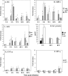
Early chemokine and cytokine expression in response to B. pertussis WT and ΔPT infection. (A to C, E, and F) Kinetics of gene expression (measured by real-time reverse transcription-PCR) of the indicated chemokines and cytokines for 48 h after infection with 5 × 10⁵ CFU of the B. pertussis WT or ΔPT strain (or after infection with PT* at selected time points). The data are mean increases relative to samples from PBS control-treated mice. (D) Levels of KC protein (measured by ELISA) in lung homogenates of mice for 48 h after infection with 5 × 10⁵ CFU of the B. pertussis WT or ΔPT strain (or PBS control-treated mice). A double dagger indicates that there was a significant increase compared with the PBS control group (four mice/group). An asterisk indicates that the P value for a comparison with the ΔPT group was <0.02.
FIG. 4.
FIG. 4.
Effect of addition of exogenous KC on recruitment of neutrophils to the airways in response to B. pertussis infection. The numbers of neutrophils in BAL fluid on days 1 and 2 after infection with 5 × 10⁵ CFU of the B. pertussis WT or ΔPT strain with intranasal administration of 1 μg KC (or PBS in control mice) 6 h postinfection (three mice/group) were determined. The asterisks indicate P values for comparisons with the ΔPT control group (*, P < 0.05; **, P < 0.01). The data are representative of the results of two similar experiments. D1, day 1; D2, day 2.
FIG. 5.
FIG. 5.
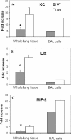
Effect of PT on expression of chemokine genes by alveolar macrophages (BAL cells) in response to B. pertussis infection in mice. Levels of gene expression (measured by real-time reverse transcription-PCR) of the indicated chemokines at 6 h after infection with 5 × 10⁵ CFU of the B. pertussis WT or ΔPT strain in whole lung tissue or BAL cells were determined. The levels of expression of KC, LIX, and MIP-2 were all greater than the control levels in lung tissues in response to WT and ΔPT infection (P < 0.05). The data are mean increases (whole lung tissue; n = 4) or increases for pooled samples from 10 mice (BAL cells) compared to samples from PBS control-treated mice. An asterisk indicates that the P value for a comparison with the ΔPT group was <0.02.
FIG. 6.
FIG. 6.
Expression of chemokine genes by MH-S cells in response to WT and ΔPT infection. The levels of KC (A), LIX (B), and MIP-2 (C) mRNA expression were significantly lower in response to WT infection than in response to ΔPT infection at an MOI of 1 at 4 and 6 h postinfection. However, at an MOI of 10, KC (D), LIX (E), and MIP-2 (F) gene expression in response to B. pertussis infection was not inhibited by PT at 4 and 6 h postinfection, suggesting that high bacterial loads may overcome the inhibitory effect of PT. A diamond indicates that the level of gene expression was not significantly greater than the control level (three wells/treatment group). The data are the results of one of two separate experiments. *, P < 0.05; **, P < 0.01.
FIG. 7.
FIG. 7.
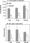
Effect of PT on neutrophil recruitment and KC gene expression in response to LPS treatment in mice. (A) Number of neutrophils in BAL fluid in mice 24 h after intranasal administration of 1, 10, and 100 ng of E. coli LPS with or without 100 ng PT. (B) KC gene expression in lungs of mice 24 h after intranasal administration of 1, 10, and 100 ng of E. coli LPS with or without 100 ng PT. A diamond indicates that the level of gene expression was not significantly greater than the control level (four mice/treatment group). The asterisks indicate P values for comparisons with the group that received only LPS (*, P < 0.03; **, P < 0.001).

Similar articles

Cited by

References

    1. Ausiello, C. M., F. Urbani, A. La Sala, R. Lande, A. Piscitelli, and A. Cassone. 1997. Acellular vaccines induce cell-mediated immunity to Bordetella pertussis antigens in infants undergoing primary vaccination against pertussis. Dev. Biol. Stand. 89315-320. - PubMed
    1. Barnard, A., B. P. Mahon, J. Watkins, K. Redhead, and K. H. Mills. 1996. Th1/Th2 cell dichotomy in acquired immunity to Bordetella pertussis: variables in the in vivo priming and in vitro cytokine detection techniques affect the classification of T-cell subsets as Th1, Th2 or Th0. Immunology 87372-380. - PMC - PubMed
    1. Brito, G. A., M. H. Souza, A. A. Melo-Filho, E. L. Hewlett, A. A. Lima, C. A. Flores, and R. A. Ribeiro. 1997. Role of pertussis toxin A subunit in neutrophil migration and vascular permeability. Infect. Immun. 651114-1118. - PMC - PubMed
    1. Carbonetti, N. H. 2007. Immunomodulation in the pathogenesis of Bordetella pertussis infection and disease. Curr. Opin. Pharmacol. 7272-278. - PubMed
    1. Carbonetti, N. H., G. V. Artamonova, C. Andreasen, and N. Bushar. 2005. Pertussis toxin and adenylate cyclase toxin provide a one-two punch for establishment of Bordetella pertussis infection of the respiratory tract. Infect. Immun. 732698-2703. - PMC - PubMed

Publication types

MeSH terms