. 2008 Nov;76(11):5357-65.
doi: 10.1128/IAI.00221-08. Epub 2008 Sep 2.

Dynamin-2-dependent targeting of mannheimia haemolytica leukotoxin to mitochondrial cyclophilin D in bovine lymphoblastoid cells

Affiliations

Dynamin-2-dependent targeting of mannheimia haemolytica leukotoxin to mitochondrial cyclophilin D in bovine lymphoblastoid cells

Dhammika N Atapattu et al. Infect Immun. 2008 Nov.

Abstract

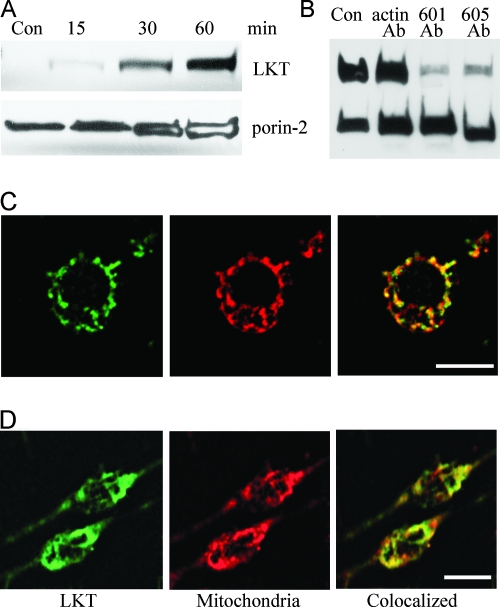
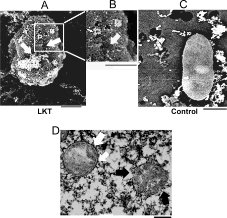
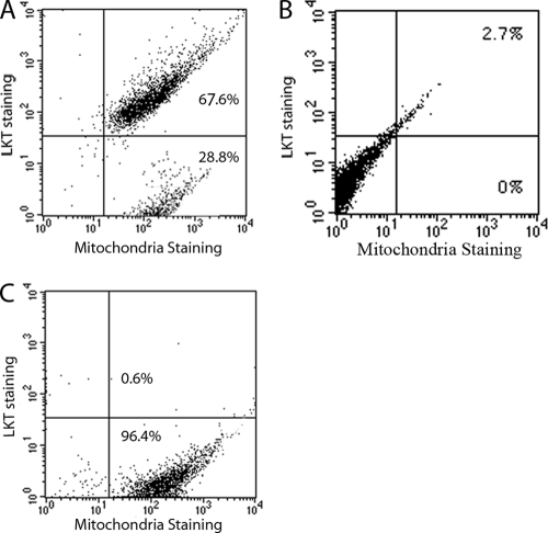
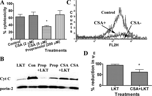
Exotoxins which belong to the family containing the RTX toxins (repeats in toxin) contribute to a variety of important human and animal diseases. One example of such a toxin is the potent leukotoxin (LKT) produced by the bovine respiratory pathogen Mannheimia haemolytica. LKT binds to CD18, resulting in the death of bovine leukocytes. In this study, we showed that internalized LKT binds to the outer mitochondrial membrane, which results in the release of cytochrome c and collapse of the mitochondrial membrane potential (psi(m)). Incubation of bovine lymphoblastoid cells (BL-3 cells) with the mitochondrial membrane-stabilizing agent cyclosporine (CSA) reduced LKT-mediated cytotoxicity, cytochrome c release, and collapse of the psi(m). Coimmunoprecipitation and intracellular binding studies suggested that LKT binds to the mitochondrial matrix protein cyclophilin D. We also demonstrated that LKT mobilizes the vesicle scission protein dynamin-2 from mitochondria to the cell membrane. Incubation with CSA depleted mitochondrial dynamin-2 in BL-3 cells, making it unavailable for vesicle scission and LKT internalization. The results of this study show that LKT trafficking and LKT-mediated cell death involve dynamin-2 and cyclophilin D, in a process that can be prevented by the mitochondrial membrane-protecting function of CSA.


Figures

FIG. 1.
FIG. 1.
LKT binds to mitochondria from BL-3 cells and RAW 264.7 cells. (A) Immunopurified mitochondria from 106 BL-3 cells were incubated for 30 to 60 min with LKT (0.5 U). Lysates were prepared and immunoblotted for LKT and the mitochondrion-specific porin-2 protein. Con, control. (B) BL-3 cells were protein transfected with anti-LKT MAbs (5 μg of MM601 or MM605) or with anti-β-actin MAb as a negative control using the Pro-ject protein transfection reagent. The cells were then incubated with LKT (0.5 U) for 1 h. Mitochondria were immunopurified and lysed, and the lysates were immunoblotted for LKT and porin-2. (C) Single BL-3 cell stained for LKT (green), mitochondria (red), and colocalization of LKT with mitochondria (yellow). (D) RAW 264.7 cells were protein transfected with LKT (0.5 U) using the Pro-ject protein transfection reagent and stained for LKT (green) and mitochondria (red) as described above. Colocalization of LKT with mitochondria is indicated by yellow. Bars = 10 μm.
FIG. 2.
FIG. 2.
LKT on mitochondria isolated from BL-3 cells detected by scanning electron microscopy and transmission electron microscopy. Mitochondria isolated from BL-3 cells were incubated with 0.5 U LKT (A and B) or RPMI medium (control) (C), stained with anti-LKT colloidal gold, and visualized by scanning electron microscopy. The micrographs show areas of LKT binding (clusters of gold beads in panels A and B) and sloughing of the MOM, leaving punched-out areas (arrows in panels A and B). (D) Mitochondrion with a disrupted outer membrane and cristae (black arrows) and normal mitochondrion with an intact double membrane (white arrow). Bars = 200 nm.
FIG. 3.
FIG. 3.
LKT detected by flow cytometry of purified mitochondria from LKT-treated BL-3 cells. Mitochondria were stained with Mitotracker Red and FITC-conjugated anti-LKT MAb. (A) Mitochondria with positive staining for both LKT and Mitotracker Red. (B and C) Mitochondria from BL-3 cells not treated with LKT (B) and mitochondria from LKT-treated cells stained with Mitotracker Red and an isotype-matched primary Ab as controls (C). The data represent the results of three similar experiments that were performed.
FIG. 4.
FIG. 4.
CSA protects BL-3 cells against LKT-mediated cytotoxicity, mitochondrial membrane damage, and collapse of ψm. (A) BL-3 cells (106 cells) were preincubated with CSA (2 or 5 μM) for 30 min before incubation with LKT (0.5 U) for 60 min. Cytotoxicity was measured using the Cell Titre 96 AQ one-assay system. (B) Mitochondria were immunopurified from untreated BL-3 cells, preincubated with CSA (5 μM) or with propranolol (Prop) (200 μM), and then incubated with LKT (0.2 U) for 30 min at 37°C. Immunoblot analysis of the resulting mitochondrial lysates demonstrated that CSA, but not propranolol, protected against LKT-mediated mitochondrial cytochrome c (Cyt c) release. Con, control. (C and D) BL-3 cells were preincubated with CSA or medium (control) and then incubated with LKT (0.5 U) for 1 h. Cells were then incubated with JC-1, a ψm-sensitive dye, at 37°C for 15 min and washed three times in PBS. Aggregated intramitochondrial JC-1 is red, and the monomeric cytoplasmic form is green. BL-3 cells were then analyzed by flow cytometry in the FL2 channel (absorption and emission maxima at 580 and 595 nm, respectively). The results show that CSA protected against collapse of ψm. The bars in panel D indicate the means of three separate experiments, and the error bars indicate the standard errors of the means. *, P < 0.05.
FIG. 5.
FIG. 5.
Movement of dynamin-2 to the cell periphery in LKT-treated BL-3 cells. (A) BL-3 cells (106 cells) were incubated with LKT (0.5 U) for 30 or 60 min and then fixed, permeabilized, stained for cytoplasmic dynamin-2 (arrows), and visualized by confocal microscopy. (B) BL-3 cells were incubated with CSA (5 μM for 30 or 60 min at 37°C). Mitochondria were isolated and lysed, and the lysates were immunoblotted for dynamin-2 and porin-2. (C) BL-3 cells preincubated with CSA were treated with LKT for 30 or 60 min. Their mitochondria were then isolated and lysed, and the lysates were immunoblotted for LKT, dynamin-2, and porin-2. (D) Mitochondrial (Mito) dynamin-2 and LKT levels in immunoblots as determined by densitometry. The bars indicate the means of three separate experiments, and the error bars indicate the standard errors of the means (*, P < 0.05). OD, optical density.
FIG. 6.
FIG. 6.
Dynamin-2 and β-actin are required for targeting of LKT to mitochondria. (A) Representative immunoblot of cell lysates from BL-3 cells transfected with dynamin-2 siRNA, untreated cells (control [Con cells]), or cells transfected with scrambled siRNA (Con siRNA). (B) BL-3 cells were transfected with dynamin-2 siRNA or scrambled siRNA or were not treated (Con Cells) before incubation with LKT (0.5 U) at 37°C for 1 h. Mitochondria were purified and lysed, and the lysates were immunoblotted for LKT and porin-2 (mitochondrial marker). (C) BL-3 cells were pretreated with cytochalasin D (CytD) (2 μg/ml for 1 h at 37°C) before they were treated with LKT. Mitochondria were then isolated and immunoblotted for LKT and porin-2. The controls included cells treated with cytochalasin D alone and untreated BL-3 cells.
FIG. 7.
FIG. 7.
LKT binds to the mitochondrial matrix protein cyclophilin D. (A) BL-3 cells were protein transfected with anti-cyclophilin D Ab (5 and 10 μg) and then incubated with LKT (0.5 U for 1 h at 37°C), and cytotoxicity was measured as described previously. BL-3 cells transfected with anti-cyclophilin D Ab (10 μg) alone or LKT-treated untransfected cells served as controls. (B) BL-3 cells were protein transfected with anti-cyclophilin D Ab (5 or 10 μg) or anti-β-actin MAb (unrelated Ab control) and then incubated with LKT (0.5 U for 1 h at 37°C). BL-3 cells incubated with LKT alone (Con LKT) and untreated BL-3 cells served as controls. Mitochondria were then purified, lysed, and immunoblotted for LKT and porin-2. (C) BL-3 cells were protein transfected with anti-cyclophilin D Ab or anti-β-actin MAb (unrelated control Ab) and incubated with LKT (0.5 U for 1 h at 37°C). Mitochondria were isolated and lysed, and the lysates were immunoblotted for cytochrome c (Cyt C) and porin-2. (D) LKT was bound to agarose protein A/G beads coated with anti-LKT Ab and incubated overnight at 4°C with a preabsorbed mitochondrial lysate (Mito lysate) from untreated BL-3 cells. The beads were then washed five times in 0.05% Triton X-100-PBS wash buffer, and proteins were eluted with Laemmli buffer. The eluates were immunoblotted for cyclophilin D and LKT. LKT bound to agarose protein A/G beads alone and mitochondrial lysate from untreated BL-3 cells served as controls. (E) Immunoblot demonstrating that preincubation with CSA (5 μM for 30 min at 37°C) reduced binding of LKT (0.5 U for 30 min at 37°C) to purified mitochondria from untreated BL-3 cells.

References

    1. Ambagala, T. C., A. P. Ambagala, and S. Srikumaran. 1999. The leukotoxin of Pasteurella haemolytica binds to beta(2) integrins on bovine leukocytes. FEMS Microbiol. Lett. 179161-167. - PubMed
    1. Arnoult, D., B. Gaume, M. Karbowski, J. C. Sharpe, F. Cecconi, and R. J. Youle. 2003. Mitochondrial release of AIF and EndoG requires caspase activation downstream of Bax/Bak-mediated permeabilization. EMBO J. 224385-4399. - PMC - PubMed
    1. Atapattu, D. N., and C. J. Czuprynski. 2007. Mannheimia haemolytica leukotoxin binds to lipid rafts in bovine lymphoblastoid cells and is internalized in a dynamin-2- and clathrin-dependent manner. Infect. Immun. 754719-4727. - PMC - PubMed
    1. Atapattu, D. N., and C. J. Czuprynski. 2005. Mannheimia haemolytica leukotoxin induces apoptosis of bovine lymphoblastoid cells (BL-3) via a caspase-9-dependent mitochondrial pathway. Infect. Immun. 735504-5513. - PMC - PubMed
    1. Battino, M., S. Bompadre, L. Leone, A. Pugnaloni, C. Rubini, M. S. Ferreiro, I. Gallardo, and P. Bullon. 2003. The effect of cyclosporine A chronic administration on the antioxidant pattern of rat liver mitochondria: structural and functional consequences. Biofactors 18271-275. - PubMed

Publication types

MeSH terms

LinkOut - more resources