. 2008 Nov;76(11):4883-94.
doi: 10.1128/IAI.01288-07. Epub 2008 Sep 2.

The gamma interferon (IFN-gamma)-inducible GTP-binding protein IGTP is necessary for toxoplasma vacuolar disruption and induces parasite egression in IFN-gamma-stimulated astrocytes

Affiliations

The gamma interferon (IFN-gamma)-inducible GTP-binding protein IGTP is necessary for toxoplasma vacuolar disruption and induces parasite egression in IFN-gamma-stimulated astrocytes

T Melzer et al. Infect Immun. 2008 Nov.

Abstract

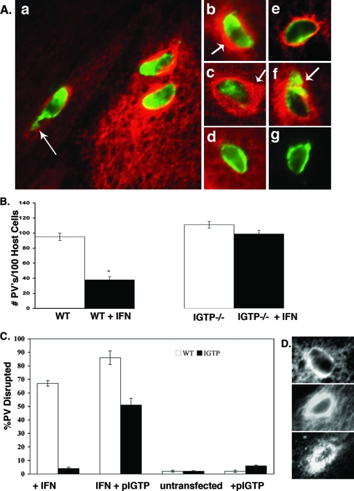
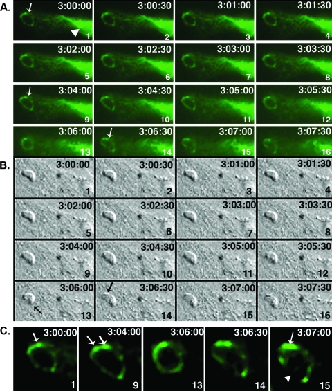
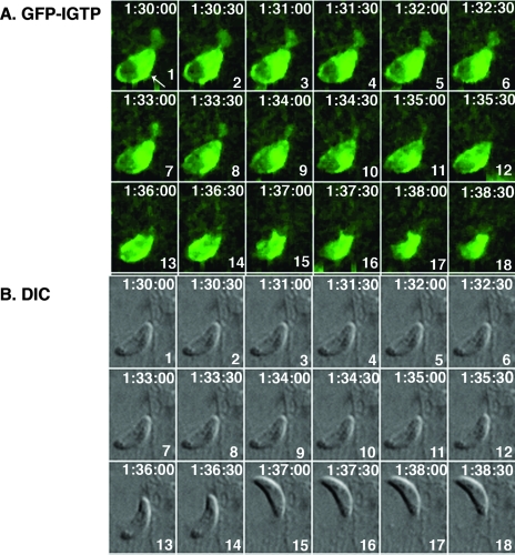
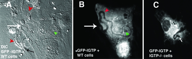
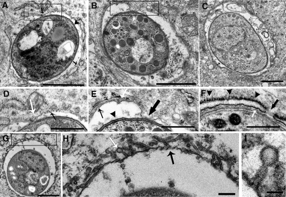
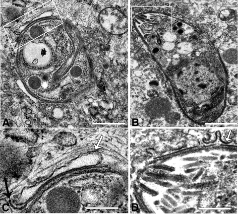
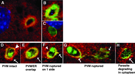
Toxoplasma gondii is a common central nervous system infection in individuals with immunocompromised immune systems, such as AIDS patients. Gamma interferon (IFN-gamma) is the main cytokine mediating protection against T. gondii. Our previous studies found that IFN-gamma significantly inhibits T. gondii in astrocytes via an IFN-gamma-inducible GTP-binding protein (IGTP)-dependent mechanism. The IGTP-dependent-, IFN-gamma-stimulated inhibition is not understood, but recent studies found that IGTP induces disruption of the parasitophorous vacuole (PV) in macrophages. In the current study, we have further investigated the mechanism of IFN-gamma inhibition and the role of IGTP in the vacuolar disruption in murine astrocytes. Vacuolar disruption was found to be dependent upon IGTP, as PV disruption was not observed in IGTP-deficient (IGTP(-/-)) astrocytes and PV disruption could be induced in IGTP(-/-) astrocytes transfected with IGTP. Live-cell imaging studies using green fluorescent protein-IGTP found that IGTP is delivered to the PV via the host cell endoplasmic reticulum (ER) early after invasion and that IGTP condenses into vesicle-like structures on the vacuole just prior to PV disruption, suggesting that IGTP is involved in PV disruption. Intravacuolar movement of the parasite occurred just prior to PV disruption. In some instances, IFN-gamma induced parasite egression. Electron microscopy and immunofluorescence studies indicate that the host cell ER fuses with the PV prior to vacuolar disruption. On the basis of these results, we postulate a mechanism by which ER/PV fusion is a crucial event in PV disruption. Fusion of the ER with the PV, releasing calcium into the vacuole, may also be the mechanism by which intravacuolar parasite movement and IFN-gamma-induced parasite egression occur.


Figures

FIG. 1.
FIG. 1.
IGTP accumulates around PVs and is necessary for vacuolar disruption in astrocytes. (A) Immunofluorescence of IGTP association with the PV in IFN-γ-stimulated WT astrocytes. Cells were stained with anti-Toxoplasma polyclonal antibody (green), anti-IGTP (red) in panels a to d, and anti-Gra7 (green) and anti-IGTP (red) in panels e to g. (a) Low-magnification view of IFN-γ-stimulated astrocytes with three disrupted vacuoles showing terminal perturbation of vacuoles (arrow), typical of a disrupted vacuole. (b) Vacuole at 2 h postinvasion showing dense circumferential layer of IGTP (arrow). (c) Confocal microscopy of vacuole at 2 h postinvasion showing the cytoplasmic layer of IGTP around the vacuole and some colocalization of IGTP and parasite antigen; note the asymmetric concentration of IGTP on one side of the vacuole (arrow). (d) Vacuole containing heat-killed Toxoplasma parasites in IFN-γ-stimulated cells. (e) Vacuole stained with Gra7 and IGTP in IFN-γ-stimulated cells at 30 min postinvasion; note the accumulation of IGTP at the vacuole. (f) Vacuole stained with Gra7 and IGTP at 2 h postinvasion, with the arrow indicating the Gra7 staining typical of disrupted vacuoles. (g) Vacuole stained with Gra7 in unstimulated cells. (B) Loss of PVs in untreated versus IFN-γ-stimulated WT and IGTP−/− astrocytes. Astrocytes were stimulated with IFN-γ for 72 h, infected with T. gondii, fixed 2 h later, stained for IGTP, and assessed for vacuolar disruption. In some groups, cells were transfected with GFP-IGTP plasmid only (+pIGTP) or with IFN-γ stimulation and the GFP-IGTP plasmid (IFN + pIGTP) and then infected, fixed 2 h later, stained for IGTP, and assessed for vacuolar disruption. In the transfected IGTP−/− cells, vacuolar disruption was assessed only for those IGTP−/− cells positive for GFP-IGTP; 50 to 100 vacuoles from each replicate were counted. Shown are the mean values for two independent experiments. * denotes that the means are statistically significant (P < 0.05; Student's t test). (D) Comparison of IGTP accumulation around the vacuole in IFN-γ-stimulated, IGTP-transfected WT, and IGTP−/− cells. Shown are IFN-γ-stimulated WT astrocytes (top); IGTP-transfected, IFN-γ-stimulated WT astrocytes (middle); and IGTP-transfected, IFN-γ-stimulated IGTP−/− astrocytes (bottom). All cells were stimulated with IFN-γ, infected with T. gondii, fixed, and stained for IGTP.
FIG. 2.
FIG. 2.
Live-cell imaging of GFP-IGTP interaction with the vacuole. (A and B) Time lapse series of GFP-IGTP (A) and the corresponding DIC images (B) beginning at 3 h postinvasion. The elapsed time (h:min:s) between each frame is shown in the upper right hand corner and each frame numbered in the lower right hand corner. Note that at 3 h postinvasion IGTP is present around the entire circumference of the PV (A, panel 1, arrow) and IGTP moves over the vacuole continuously from the host cell ER (A, panel 1, arrowhead). During the next 7 to 8 min, the intravacuolar parasite flexes twice; the first flexion occurs approximately 3 h and 3 min postinvasion (A and B, frame 8) and the second flexion 2 min later (A and B, frame 13, arrow), followed by fusion of IGTP-positive vesicles with the vacuole in discrete areas (A and B, frames 14). Moments later, the vacuole was disrupted. (C) Selected panels showing IGTP interaction with the vacuole just prior to PV disruption. IGTP is initially present on the vacuole, with a concentration at the upper edge of the vacuole (frame 1). After the first flexion, IGTP condenses into two discrete areas (frame 9, arrows), the “death flex” (frame 13) and the frames just prior to PV disruption (frames 14 and 15). Note that in frame 15 IGTP is concentrated on the upper edge of the vacuole (arrow) while IGTP is now absent from the lower edge (arrowhead), where the PV disrupts.
FIG. 3.
FIG. 3.
GFP-IGTP live-cell time series of egressing Toxoplasma parasites at 1.5 h postinvasion. (A) GFP-IGTP series and (B) the corresponding DIC images, beginning at 1.5 h postinvasion, with the elapsed time recorded as h:min:s in the upper right hand corner and the frame number in the lower right hand corner of each frame. Note that the vacuole is surrounded by IGTP, with a concentration of IGTP on the lower right side (A, frame 1, arrow). The parasite begins to egress approximately 6 min later (frames 13 and 14) and has fully exited the host cell by frame 15; note that IGTP remains around the now-empty vacuole.
FIG. 4.
FIG. 4.
IFN-γ-induced Toxoplasma egression in astrocytes. (A) Differential contrast microscopy image of live-cell imaging with GFP-IGTP-transfected WT astrocytes, with the large white arrow indicating the site of parasite egress, the black arrow the extracellular egressed parasite, the green arrowhead the original site of the intracellular vacuole, and the red arrowheads the intracellular and extracellular trails left by the egressing parasite. (B) Corresponding fluorescent image of GFP-IGTP, with the green arrowhead indicating the original site of intracellular vacuole and the red arrowhead the intracellular trail emanating from the vacuole. (C) Fluorescent image of GFP-IGTP-transfected IGTP−/− astrocytes; the white arrow indicates the site of the vacuole.
FIG. 5.
FIG. 5.
Transmission electron microscopy of the early events in PV disruption in IFN-γ-stimulated astrocytes. Shown are various views of aberrations to the vacuole and the PVM observed during the first 2 h postinvasion. (A) Vacuole at 30 min postinvasion, with point of contact between the PVM and the parasite plasma membrane (box) and invaginations of the PVM (box, black arrow); the arrowhead points to the parasite plasma membrane, and the arrow points to the PVM. Bar = 1 μm. (B) Vacuole at 2 h postinvasion, with enlarged area of contact between the parasite plasma membrane and the PVM (box). Bar = 1 μm. (C) Vacuole in unstimulated astrocyte, showing the normal arrangement of the PVM and ER. Bar = 0.5 μm. (D) High-magnification view of the area of contact outlined in the box in panel A; note the area of contact between the PVM and the parasite (white arrow) and the invaginations of the PVM (black arrow). Bar = 0.5 μm. (E) High-magnification view of the boxed area in panel B; note the area of contact between the PVM and the parasite (large black arrow) and the disrupted ER (white arrow); the parasite plasma membrane is indicated by the black arrowhead and the PVM by the small black arrow. Bar = 0.5 μm. (F) High-magnification view of vacuole showing apparent dissolution of the PVM at the area between the folds of the ER (black arrow) and the close opposition of the ER and PVM (white arrow); black arrowheads indicate the layer of rough ER surrounding the PV. Bar = 0.5 μm. (G) Vacuole showing small, stemmed vesicular structures fusing with the PVM. Bar = 1 μm. (H) High-magnification view of the boxed area in panel G, showing stemmed vesicular and tubular structures (white arrow) fusing with the PVM and PVM indentations (black arrow). Bar = 0.1 μm. (I) High-magnification view showing the hollow, stemmed membrane structure fusing with the vacuole. Bar = 50 nm.
FIG. 6.
FIG. 6.
Late stages in PV disruption. (A) Parasite in disrupted vacuole showing the remnant of the PVM and the delaminating parasite plasma membrane (box). Bar = 1 μm. (B) Parasite free in the cytosol, with evidence of PVM vesiculation (box). Bar = 1 μm. (C). High-magnification view of the boxed area in panel A; the white arrow indicates the PVM, and the black arrow indicates the parasite plasma membrane. Bar = 0.1 μm. (D) High-magnification view of boxed area in panel B; the arrow indicates the PVM. Bar = 0.1 μm.
FIG. 7.
FIG. 7.
Interaction of the ER with the PVM in IFN-γ-stimulated astrocytes. (A to C) Immunofluorescence microscopy of IFN-γ-stimulated astrocytes labeled with the ER membrane markers calnexin (red) and Gra7 (green) in IFN-γ-stimulated astrocytes at 30 min postinvasion (A) and 2 h postinvasion (B) and unstimulated astrocytes at 2 h postinvasion (C). All cells were counterstained with the nuclear dye DAPI. (D to H) Confocal microscopy of PVs from IFN-γ-stimulated astrocytes labeled with the ER membrane markers calnexin (red) and Gra7 (green): (D) intact PV surrounded by host ER (arrow) and with a small area of ER and Gra7 colocalization (arrowhead), (E) intact PV with a thick PVM/ER layer (arrow), (F) disrupted PV with one side devoid of Gra7 (arrowhead) and an ER/Gra7 extension on the opposite side (arrow), (G) disrupted PV with Gra7 extension into the cytoplasm (arrows), and (H) disrupted PV with degrading parasite (arrow). In unstimulated WT astrocytes, no such areas of ER/Gra7 overlap were observed (C).

Similar articles

Cited by

References

    1. Adams, L. B., J. B. Hibbs, Jr., R. R. Taintor, and J. L. Krahenbuhl. 1990. Microbiostatic effect of murine-activated macrophages for Toxoplasma gondii. Role for synthesis of inorganic nitrogen oxides from L-arginine. J. Immunol. 1442725-2729. - PubMed
    1. Arrizabalaga, G., and J. C. Boothroyd. 2004. Role of calcium during Toxoplasma gondii invasion and egress. Int. J. Parasitol. 34361-368. - PubMed
    1. Bekpen, C., J. P. Hunn, C. Rohde, I. Parvanova, L. Guethlein, D. M. Dunn, E. Glowalla, M. Leptin, and J. C. Howard. 2005. The interferon-inducible p47 (IRG) GTPases in vertebrates: loss of the cell autonomous resistance mechanism in the human lineage. Genome Biol. 6R92. - PMC - PubMed
    1. Black, M. W., and J. C. Boothroyd. 2000. Lytic cycle of Toxoplasma gondii. Microbiol. Mol. Biol. Rev. 64607-623. - PMC - PubMed
    1. Butcher, B. A., R. I. Greene, S. C. Henry, K. L. Annecharico, J. B. Weinberg, E. Y. Denkers, A. Sher, and G. A. Taylor. 2005. p47 GTPases regulate Toxoplasma gondii survival in activated macrophages. Infect. Immun. 733278-3286. - PMC - PubMed

Publication types

MeSH terms