Review
. 2008 Sep 1;22(17):2308-41.
doi: 10.1101/gad.1686208.

Deciphering the function of canonical Wnt signals in development and disease: conditional loss- and gain-of-function mutations of beta-catenin in mice

Affiliations
Review

Deciphering the function of canonical Wnt signals in development and disease: conditional loss- and gain-of-function mutations of beta-catenin in mice

Tamara Grigoryan et al. Genes Dev. .

Abstract

Wnt signaling is one of a handful of powerful signaling pathways that play crucial roles in the animal life by controlling the genetic programs of embryonic development and adult homeostasis. When disrupted, these signaling pathways cause developmental defects, or diseases, among them cancer. The gateway of the canonical Wnt pathway, which contains >100 genes, is an essential molecule called beta-catenin (Armadillo in Drosophila). Conditional loss- and gain-of-function mutations of beta-catenin in mice provided powerful tools for the functional analysis of canonical Wnt signaling in many tissues and organs. Such studies revealed roles of Wnt signaling that were previously not accessible to genetic analysis due to the early embryonic lethality of conventional beta-catenin knockout mice, as well as the redundancy of Wnt ligands, receptors, and transcription factors. Analysis of conditional beta-catenin loss- and gain-of-function mutant mice demonstrated that canonical Wnt signals control progenitor cell expansion and lineage decisions both in the early embryo and in many organs. Canonical Wnt signaling also plays important roles in the maintenance of various embryonic or adult stem cells, and as recent findings demonstrated, in cancer stem cell types. This has opened new opportunities to model numerous human diseases, which have been associated with deregulated Wnt signaling. Our review summarizes what has been learned from genetic studies of the Wnt pathway by the analysis of conditional beta-catenin loss- and gain-of-function mice.


Figures

Figure 1.
Figure 1.
Scheme of the Wnt signaling pathway. The Wnt glycoproteins are produced by the neighboring cells and secreted in a process that involves many components, such as a multiprotein retromer complex, and the multispanning transmembrane protein Wntless (Wls). The secreted Wnt ligands bind to the receptors of the seven-membrane-spanning Frizzled family and single-spanning LRP family. Secreted factors like secreted Frizzled-related proteins (sFRPs) bind to Wnts and block the interaction with Frizzled receptors. Dickkopf antagonizes Wnt signaling by blocking LRP receptors. Three main branches are activated downstream from the Wnt signals: canonical Wnt/β-catenin signaling (blue and red, in the center and on the left), and the noncanonical Jun kinase and Wnt/Ca2+ pathways (green and orange on the right). The central component of the canonical Wnt signaling is β-catenin (red). (Left and right) In the absence of the Wnt signals, β-catenin forms a stable complex with E-cadherin and participates in adherens junction formation. (Left) Excess β-catenin is captured by a destruction complex containing GSK3β, Axin1, Axin2/Conductin, and APC, where it gets N-terminally phosphorylated and, following ubiquitination by β-Trcp, targeted to proteosome-mediated degradation. In contrast, in the presence of the Wnt ligands, disassembly of the destruction complex occurs through phosphorylation of LRP5/6 by GSK3β and CK1α, and binding of Axin to LRP, thereby preventing β-catenin degradation. Free cytoplasmatic β-catenin can then enter the nucleus, where it forms a transcriptionally active complex with Tcf/Lef family of transcription factors, by replacing Grouchos from chromatin, and interacting with other coactivators like CBP, BCL9, and Pygopus. The three pathways branch at the level of Dishevelled (Dsh) protein. In the noncanonical Jun kinase pathway, Dsh via Daam1, is connected to downstream effectors, such as Rho, and regulates cytoskeletal organization and cell polarity. The Wnt/Ca2+ pathway leads to the release of intracellular calcium, and involves activation of Phospholipase C (PLC) and protein kinase C (PKC). Ca2+ can activate Calmodulin-dependent protein kinase C (CaMKII), and Nemo-like kinase (NLK), and suppress canonical Wnt signaling.
Figure 2.
Figure 2.
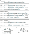
Introduction of conditional loss- and gain-of-function mutations into the β-catenin locus. (a) Genomic locus of the β-catenin gene and the two strategies of generation of the β-catenin loss-of-function alleles. In one strategy, loxP sites flank exons 3–6, to generate the Ctnnb1Wbm/flox allele; in the second strategy, loxP sites flank exons 2–6 to generate the Ctnnb1Kem/flox allele. In both cases, the floxed parts encode the N-terminal domain and the Armadillo repeats 1–4 of the β-catenin protein. Transient expression of cre recombinase results in excision of the neo resistance cassette. Subsequently, tissue-specific cre expression leads to excision of the floxed exons, frameshift mutations, and production of a null-allele (see in c, cf. wild type with LOF). (b) Generation of the β-catenin gain-of-function allele. LoxP sites flank exon 3, which encodes phosphorylation sites targeted by GSK3β. Tissue-specific cre expression leads to excision of exon 3 and in-frame splicing on exons 2 and 4, thus resulting in expression of a protein that escapes the degradation. (c,d) Western blot analysis showing tissue-specific recombination of the targeted loci. In the conditional loss-of-function mutant, no β-catenin protein can be detected, whereas in the gain-of-function mutants, a shortened β-catenin variant is produced (cf. wild type, LOF, and GOF). On the right, the expression of the mutant β-catenin protein after cre-mediated recombination in cytoplasmic (Cyt) and nuclear (Nu) fractions of the recombinant embryonic stem cells is shown. (WT) Wild type; (LOF) loss-of-function allele; (GOF) gain-of-function allele. d was reprinted with permission from Macmillan Publishers Ltd: Harada et al. (1999), © 1999; and c was reprinted from Zechner et al. (2003) with permission from Elsevier.
Figure 3.
Figure 3.
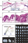
Wnt/β-catenin signaling in skin and teeth. (a,b) Conditional loss-of-function mutations of β-catenin in skin prevent hair follicle generation and result in formation of cysts at P32. Stem cells are localized to the specific areas close to the cysts in β-catenin loss-of-function mutants, as shown by in situ hybridization for the stem cell marker Keratin 15 (arrows) and immunohistochemistry for β1-integrin (insets, arrows). (c,d) Conditional gain-of-function mutations of β-catenin in skin result in disturbed hair follicle morphogenesis at E17, which is accompanied by epidermal thickening (brackets) and the formation of additional hair placodes (d, arrows and arrowheads), as well as in keratin deposits (d, asterisks). (e) Scheme of the role of β-catenin in fate decisions of the bulge stem cells toward follicular or epidermal lineages. (f,g) Conditional gain-of-function mutations of β-catenin in tooth epithelium lead to generation of supernumerary teeth of various developmental stages and sizes from one mutant molar tooth bud. a, b, and e are reprinted with permission from Huelsken et al. (2001) with permission from Elsevier. c and d are reprinted by permission of the Company of Biologists from Närhi et al. (2008). f and g are reprinted with permission from Järvinen et al. (2006); © 2006 National Academy of Sciences, U.S.A.
Figure 4.
Figure 4.
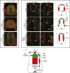
Wnt/β-catenin signaling in the spinal cord. (a–c) Changes in the size of the neural progenitor pool in β-catenin mutants. At E11.5, sizes of spinal cords and the neural progenitor pool are reduced in β-catenin loss-of-function mutants (a,b) and increased in β-catenin gain-of-function mutants, as shown by Nestin-specific antibodies in green (a,c). Accordingly, the mantle zones are increased in the loss-of-function mutants (a,b) and decreased in gain-of-function mutants (a,c), as shown by Tubulin J1-specific antibodies in red. (d–m) Changes in neuronal specification in β-catenin mutants. Differentiated neurons are shown by antibody staining against Foxd3, dI2 (darker green), Isl1/2, dI3 (light green), and Lbx1, dI4-6 (red). Arrowheads label motor neurons (M) and V1 interneurons (V1) in the ventral spinal cord, and asterisks label the dorsal root ganglia. Progenitor domains that generate dI2 and dI3 neurons are expanded in β-catenin gain-of-function mutants (cf. d,g,j and e,h,k, labeled with vertical brackets). In compound mutants that carry a gain-of-function mutation of β-catenin and loss-of-function mutations of Olig3, only Lbx1+ but not Foxd3+ and Isl1/2+ neurons are generated in the dorsal spinal cord (f,i,l), indicating requirement of Olig3 expression for dI2 and dI3 neuron generation. (m) In the neuronal progenitor cells, Wnt/β-catenin acts downstream from Bmp signaling to promote specification of dI2/3 neuronal lineages and restrict expansion of dI4-6 lineages via the patterning gene Olig3. Reprinted with permission from Zechner et al. (2003, 2007) with permission from Elsevier.
Figure 5.
Figure 5.
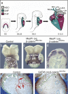
Wnt/β-catenin signaling during cardiogenesis. (a) Schemes of mouse embryos at different stages of embryonic development show the location and contribution of first (red) and second (green) heart field cells at cardiac crescent (frontal view), linear heart tube, and looping stages (lateral views). Following rightward looping of the linear heart tube, neural crest cells migrate into the outflow tract and take part in septation and remodeling of the outflow tract, resulting in a multichambered heart at E13.5. (b–d) Conditional ablation and activation of β-catenin using MesP1-driven cre shows the loss of cardiac looping in MesP1 cre; β-cateninlox/lox mice (b,c), and the disruption of cardiac tube formation in MesP1 cre, β-cateninloxEx3/+ mice (b,d) as indicated by whole-mount in situ hybridization for Nkx2.5 and MLC2a at E8.5. (e,f) Conditional ablation of β-catenin in the proepicardial lineage (Gata5 cre) resulted in the absence of coronary arterial vasculature as indicated by PECAM 1 staining at E18.5 (cf. red arrows in e with f). (Ao) Aorta; (DA) ductus arteriosus; (FHF) first heart field; (IFT) inflow tract; (LV) left ventricle; (NCC) neural crest; (OFT) outflow tract; (PA) pulmonary artery; (pV) primitive ventricle; (RV) right ventricle; (RA and LA) right and left atrium; (RCA) right carotid artery; (LCA) left carotid artery; (RSCA) right subclavian artery; (LSCA) left subclavian artery; (SHF) second heart field. b–d are reprinted with permission from Klaus et al. (2007); © 2007 National Academy of Sciences, U.S.A. e and f are reprinted with permission from Zamora et al. (2007); © 2007 National Academy of Sciences, U.S.A.
Figure 6.
Figure 6.
Wnt/β-catenin pathway directly induces Pitx2 expression in the pituitary gland. (a) LiCl addition to the primary cell culture prepared from E10.5 pituitary glands leads to activation of the Pitx2-driven LacZ expression. (b) Overexpression of the constitutively active β-catenin (ΔN-β-catenin) in the αT3-1 pituitary cell line leads to activation of Pitx2 gene expression. (c) Chromatin immunoprecipitation analysis demonstrated that in the LiCl-stimulated cells, β-catenin binds to the Pitx2 promoter together with LEF1, whereas in the LiCl-unstimulated cells, LEF1 binds to the Pitx2 promoter along with HDAC1. Reprinted from Kioussi et al. (2002) with permission from Elsevier.
Figure 7.
Figure 7.
Wnt/β-catenin signaling in somitogenesis. (a,b) Conditional loss-of-function mutations of β-catenin result in anterior mesoderm thickening, posterior truncation and prevention of somitogenesis. (a,b) Tbx6, a marker for posterior presomitic mesoderm, is lost in loss-of-function mutants. Conditional gain-of-function mutations of β-catenin result in posterior mesoderm expansion shown by expanded Tbx6 expression (a,c). (d–f) Expression of Mesp2 is lost in loss-of-function mutants (d,e), and appears in multiple stripes in gain-of-function mutants (d,f). Reprinted by permission of the Company of Biologists from Dunty et al. (2008).
Figure 8.
Figure 8.
Wnt/β-catenin signaling in limb patterning. (a) At 42 somite stage AER is absent or severely reduced in β-catenin loss-of-function mutants (β-catflox), and expanded in the gain-of-function mutants (ΔN-β-cat), as shown by Fgf8 and Bmp4 expression. AER is not formed in BmpR1a loss-of-function mutants (BmpR1Aflox) at 42 somite stage. In compound mutants with loss-of-function mutations of BmpR1a and gain-of-function mutations of β-catenin AER is expanded, resembling the phenotype of single β-catenin gain-of-function mutants. This indicates that canonical Wnt signaling acts downstream from Bmp signaling in AER formation. (b) At 30 somite stage expression of the ventral gene En1 is lost and expression of the dorsal gene Wnt7a is expanded ventrally in β-catenin loss-of-function mutants. In contrast, in gain-of-function mutants, expression of these markers is unchanged. In BmpR1a loss-of-function mutants, as well as in compound mutants, phenotypes resemble that of β-catenin loss-of-function mutants, indicating that canonical Wnt signaling acts upstream of, or in parallel to the Bmp signaling in D–V patterning of the limb. All gene mutations were introduced using Brain 4 cre. Reprinted with permission from Soshnikova et al. (2003).

References

    1. Aberle H., Butz S., Stappert J., Weissig H., Kemler R., Hoschuetzky H. Assembly of the cadherin–catenin complex in vitro with recombinant proteins. J. Cell Sci. 1994;107:3655–3663. - PubMed
    1. Abu-Elmagd M., Garcia-Morales C., Wheeler G.N. Frizzled7 mediates canonical Wnt signaling in neural crest induction. Dev. Biol. 2006;298:285–298. - PubMed
    1. Ahn K., Mishina Y., Hanks M.C., Behringer R.R., Crenshaw E.B. BMPR-IA signaling is required for the formation of the apical ectodermal ridge and dorsal–ventral patterning of the limb. Development. 2001;128:4449–4461. - PubMed
    1. Ai D., Fu X., Wang J., Lu M.F., Chen L., Baldini A., Klein W.H., Martin J.F. Canonical Wnt signaling functions in second heart field to promote right ventricular growth. Proc. Natl. Acad. Sci. 2007;104:9319–9324. - PMC - PubMed
    1. Akiyama H., Lyons J.P., Mori-Akiyama Y., Yang X., Zhang R., Zhang Z., Deng J.M., Taketo M.M., Nakamura T., Behringer R.R., et al. Interactions between Sox9 and β-catenin control chondrocyte differentiation. Genes & Dev. 2004;18:1072–1087. - PMC - PubMed

Publication types

MeSH terms

LinkOut - more resources