Comparative Study
. 2008 Sep 1;22(17):2385-99.
doi: 10.1101/gad.1687508.

A new mechanism controlling kinetochore-microtubule interactions revealed by comparison of two dynein-targeting components: SPDL-1 and the Rod/Zwilch/Zw10 complex

Affiliations
Comparative Study

A new mechanism controlling kinetochore-microtubule interactions revealed by comparison of two dynein-targeting components: SPDL-1 and the Rod/Zwilch/Zw10 complex

Reto Gassmann et al. Genes Dev. .

Abstract

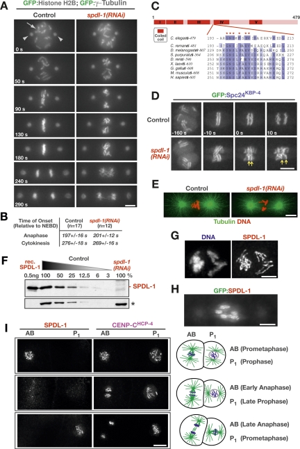
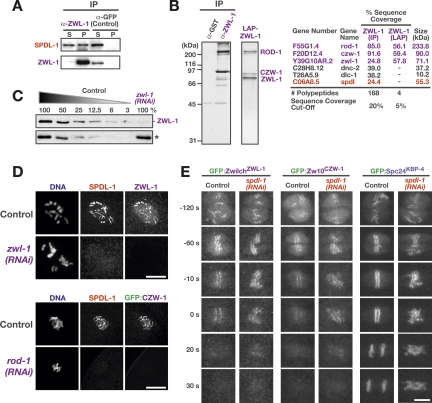
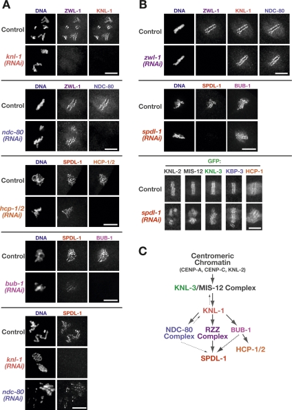
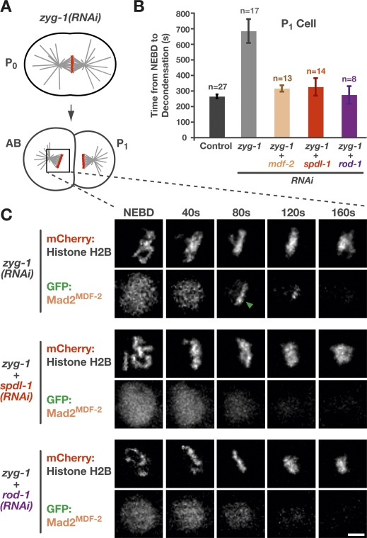
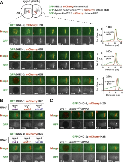
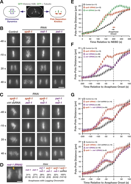
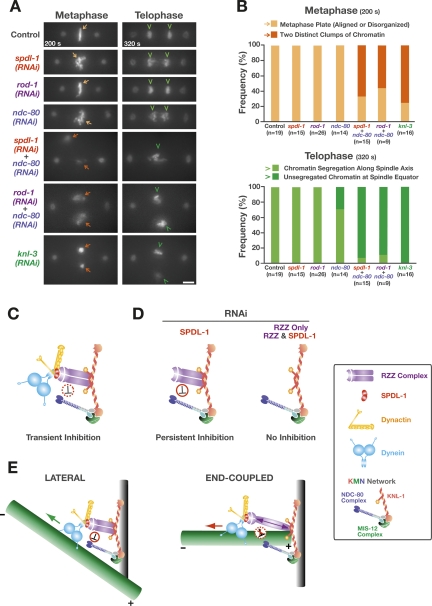
Chromosome segregation requires stable bipolar attachments of spindle microtubules to kinetochores. The dynein/dynactin motor complex localizes transiently to kinetochores and is implicated in chromosome segregation, but its role remains poorly understood. Here, we use the Caenorhabditis elegans embryo to investigate the function of kinetochore dynein by analyzing the Rod/Zwilch/Zw10 (RZZ) complex and the associated coiled-coil protein SPDL-1. Both components are essential for Mad2 targeting to kinetochores and spindle checkpoint activation. RZZ complex inhibition, which abolishes both SPDL-1 and dynein/dynactin targeting to kinetochores, slows but does not prevent the formation of load-bearing kinetochore-microtubule attachments and reduces the fidelity of chromosome segregation. Surprisingly, inhibition of SPDL-1, which abolishes dynein/dynactin targeting to kinetochores without perturbing RZZ complex localization, prevents the formation of load-bearing attachments during most of prometaphase and results in extensive chromosome missegregation. Coinhibition of SPDL-1 along with the RZZ complex reduces the phenotypic severity to that observed following RZZ complex inhibition alone. We propose that the RZZ complex can inhibit the formation of load-bearing attachments and that this activity of the RZZ complex is normally controlled by dynein/dynactin localized via SPDL-1. This mechanism could coordinate the hand-off from initial weak dynein-mediated lateral attachments, which help orient kinetochores and enhance their ability to capture microtubules, to strong end-coupled attachments that drive chromosome segregation.


Figures

Figure 1.
Figure 1.
SPDL-1 is a transient kinetochore component essential for chromosome segregation. (A) Selected frames from a live-imaging sequence of the first division in unperturbed and spdl-1(RNAi) embryos expressing GFP:histone H2B and GFP:γ-tubulin to simultaneously visualize chromosomes (arrow) and spindle poles (arrowheads), respectively (see also Supplemental Movie 1). Images are time-aligned relative to NEBD (0 sec). Bar, 5 μm. (B) Timing of anaphase onset and cytokinesis onset in unperturbed and spdl-1(RNAi) embryos. Anaphase onset was defined as the first visible sister chromatid separation (GFP:histone H2B) and cytokinesis onset by the first visible ingression of the cleavage furrow in DIC images acquired in parallel. Values represent the S.E.M with a 95% confidence interval. (C) Primary sequence features of SPDL-1 and related proteins. The highly conserved motif that defines this conserved coiled-coil protein family is depicted (see also Supplemental Fig. 1). (D) Chromosome condensation, sister centromere resolution, and the separation of sister chromatids at anaphase onset are normal in spdl-1(RNAi) embryos. Selected frames of a live-imaging sequence are shown (see also Supplemental Movie 2). Kinetochores are marked by GFP:Spc24KBP-4, a subunit of the NDC-80 complex. Arrows highlight separating sister kinetochores at anaphase onset (0 sec) in spdl-1(RNAi) embryos. Bar, 5 μm. (E) Mitotic spindle morphology in control and spdl-1(RNAi) embryos fixed and stained with a fluorescently labeled antibody against α-tubulin (see also Supplemental Movie 3). Bar, 5 μm. (F) Immunoblotting with an affinity-purified polyclonal antibody raised against SPDL-1 detects purified recombinant (rec.) SPDL-1 and a protein of equal size in wild-type (N2) worms, which is depleted >95% by RNAi. The relative amount of worm extract loaded is indicated above each lane. A cross-reacting protein band (*) serves as the loading control. (G) Immunofluorescence image of a one-cell embryo at prometaphase immunostained for SPDL-1. Bar, 2 μm. (H) Snapshot of a one-cell embryo in prometaphase expressing GFP:SPDL-1. Bar, 5 μm. (I) SPDL-1 localizes transiently to kinetochores from prometaphase to anaphase onset. Two-cell embryos at different stages are shown costained for CENP-CHCP-4, which is present at kinetochores throughout mitosis, and SPDL-1. The natural difference in cell cycle timing of the AB and P1 cells (with AB entering and exiting mitosis prior to P1, as diagrammed on the right) defines the transient period of SPDL-1 kinetochore localization (see also Supplemental Movie 4). Bar, 5 μm.
Figure 2.
Figure 2.
SPDL-1 is recruited to the kinetochore by the RZZ complex. (A) SPDL-1 coimmunoprecipitates with ZwilchZWL-1. Worm extracts were depleted of ZwilchZWL-1 using an affinity-purified polyclonal antibody, and the resulting supernatant (S) and pellet (P) was analyzed by immunoblot. Loading of pellet is 20× relative to supernatant. An antibody against GFP was used in the control immunoprecipitation experiment. (B) SPDL-1 associates with the RZZ complex but is not a core subunit. A one-step immunoprecipitation of ZwilchZWL-1 and a stringent two-step isolation of GFPLAP-tagged ZwilchZWL-1 were visualized on a silver-stained gel and analyzed by mass spectrometry as shown on the right. (C) Immunoblotting with the anti-ZwilchZWL-1 antibody detects a 70-kDa band, which is depleted >95% following RNAi. A cross-reacting protein band (*) serves as the loading control. (D) Immunofluorescence images of early embryos stained for SPDL-1 after depletion of ZwilchZWL-1 or ROD-1. Bars, 5 μm. (E) Depletion of SPDL-1 affects neither kinetochore targeting of RZZ subunits nor their rapid disappearance from kinetochores at anaphase onset. Selected frames from time-lapse sequences of embryos expressing GFP:ZwilchZWL-1, GFP:Zw10CZW-1, and GFP:Spc24KBP-4 are shown (see also Supplemental Movies 5, 6). Time is relative to the onset of sister chromatid separation. Bar, 5 μm.
Figure 3.
Figure 3.
SPDL-1 and the RZZ complex are dispensable for the formation of the core kinetochore microtubule attachment site. (A) Consequences of outer kinetochore component depletion on the localization of SPDL-1 and ZwilchZWL-1, assayed by immunofluorescence. Bars, 5 μm. (B) Normal localization of outer kinetochore components after depletion of SPDL-1 and ZwilchZWL-1, assayed by immunofluorescence or live imaging of previously characterized GFP-fusions (Cheeseman et al. 2004, 2005; Maddox et al. 2007). Bars, 5 μm. (C) Summary of the dependency analysis for kinetochore targeting of SPDL-1 and the RZZ complex. For each depletion-localization experiment, between five and 10 one-cell or two-cell embryos were examined.
Figure 4.
Figure 4.
SPDL-1 is required for a functional spindle checkpoint and kinetochore localization of Mad2MDF-2. (A) Perturbation to generate monopolar spindles in the second division and trigger spindle checkpoint activation in C. elegans embryos. ZYG-1 is a kinase required for centriole duplication (O’Connell et al. 2001). In zyg-1(RNAi) embryos, the first division is normal, because two intact centrioles are contributed by sperm that is not affected by RNAi. These centrioles are unable to duplicate, however, resulting in a monopolar spindle in the subsequent division. (B) Average time from NEBD to chromosome decondensation in the P1 cell of a worm strain expressing GFP:histone H2B. ZYG-1 single depletion results in a significant delay that depends on Mad2MDF-2, SPDL-1, and ROD-1. Error bars represent the SEM with a 95% confidence interval. A similar result is observed in the AB cell (data not shown). (C) Stills from a time-lapse sequence of the AB cell monopolar division in a worm strain coexpressing GFP:Mad2MDF-2 and mCherry:histone H2B. In ZYG-1 single depletions, GFP:Mad2MDF-2 accumulates on kinetochores that are distal to the pole (arrow). Codepletion of ZYG-1 with SPDL-1 or ROD-1 prevents kinetochore accumulation of GFP:Mad2MDF-2 (see also Supplemental Movie 7). Bar, 5 μm.
Figure 5.
Figure 5.
Localization of dynein and dynactin to kinetochores requires SPDL-1. (A) Unattached kinetochores on second-division monopolar spindles accumulate dynein and dynactin. Strains stably coexpressing GFP:fusions of either KNL-2, full-length dynein heavy chainDHC-1, or dynamitinDNC-2 with mCherry:histone H2B were used to monitor kinetochore localization (see also Supplemental Movie 8). All images are of the AB cell, and the single spindle pole is always to the left. Times are relative to NEBD. Line scans (5 pixels wide; normalized relative to maximum intensity in each channel) indicate the bilaterally symmetric distribution of KNL-2 relative to mCherry:histone H2B, which contrasts with the asymmetric enrichment of DHC-1 and DNC-2 on the chromosomal face pointing away from the single pole. Bars, 5 μm. (B) Kinetochore accumulation of GFP:dynein heavy chainDHC-1 and GFP:dynamitinDNC-2 requires SPDL-1 and the RZZ complex but not NDC-80. For brevity, a single frame is shown for each condition (see also Supplemental Movies 9, 10). Bars, 5 μm. (C) Abrogating the spindle checkpoint by depleting Mad2MDF-2 does not prevent recruitment of GFP:dynein heavy chainDHC-1 or GFP:dynamitinDNC-2 to unattached kinetochores. However, accumulation of the GFP:fusion proteins is limited because of premature mitotic exit. Bars, 5 μm.
Figure 6.
Figure 6.
Depletion of SPDL-1 results in a more severe chromosome segregation defect than depletion of ROD-1 or ZwilchZWL-1. (A) Cartoon outlining the two parameters monitored in a strain expressing GFP:histone H2B and GFP:γ-tubulin: chromosome dynamics and kinetics of spindle pole separation. (B) Frames from time-lapse sequences of the first embryonic division, highlighting the differences in chromosome dynamics after depletion of SPDL-1 and the RZZ complex subunits ROD-1 and ZwilchZWL-1 (see also Supplemental Movie 11). The time point 0 sec denotes the onset of sister chromatid separation. Bar, 5 μm. (C) Selected frames from time-lapse sequences of embryos codepleted of RZZ subunits and SPDL-1, which significantly reduces the severe chromosome missegregation phenotype of SPDL-1 single depletions to match that of RZZ subunit single depletions (see also Supplemental Movie 12). Bar, 5 μm. (D) Representative image of anaphase with lagging chromatin in a rod-1(RNAi) one-cell embryo. The frequency of one-cell embryos with lagging anaphase chromatin is indicated for single and double inhibitions involving RZZ subunits and SPDL-1. Bar, 2 μm. (E) Pole separation kinetics in wild-type, spdl-1(RNAi), and knl-3(RNAi) embryos. Images were acquired at 10-sec intervals, and sequences were time-aligned relative to NEBD. Pole–pole distances in the time-aligned sequences were measured, averaged for the indicated number (n) of embryos, and plotted against time. Error bars represent the SEM with a 95% confidence interval. (F) Pole separation kinetics of the perturbations shown in B. Sequences were time-aligned relative to the onset of sister chromatid separation (“Anaphase Onset”). Error bars represent the SEM with a 95% confidence interval. (G) Pole separation kinetics of the perturbations shown in C, demonstrating that double depletions of SPDL-1 and RZZ complex subunits result in a pole separation profile that is indistinguishable from RZZ subunit single depletions. For controls, spdl-1 dsRNA was diluted equally with dsRNA corresponding to the budding yeast gene CTF13 or the C. elegans gene sas-5, which is not required for the first embryonic division (both conditions gave identical results). Error bars represent the SEM with a 95% confidence interval.
Figure 7.
Figure 7.
Codepletion of SPDL-1 or ROD-1 with NDC-80 recapitulates the “kinetochore-null” phenotype. (A) Frames from time-lapse sequences representing metaphase (200 sec after NEBD) and telophase (320 sec after NEBD). Codepletion of SPDL-1 or ROD-1 with NDC-80 approximates the “kinetochore-null” phenotype of knl-3(RNAi) embryos (see also Supplemental Movies 13 and 14), in which chromosomes of the two pronuclei are often visible as separate clumps at metaphase (arrows), and unsegregated chromatin remains at the spindle equator in telophase (arrowheads). Bar, 5 μm. (B) Percentage of first divisions displaying the chromosome morphologies described in A at 200 sec and 320 sec after NEBD. (C) Schematic summary of the relationship between the RZZ complex, SPDL-1, dynein/dynactin, and the NDC-80 complex. The negative regulation of the KMN network by the RZZ complex, which is transient in the wild-type situation, may be either direct or indirect. (D) Model explaining the difference in phenotypic severity between SPDL-1 and RZZ complex inhibitions. Specifically, we propose that SPDL-1 depletion results in persistent RZZ complex-mediated inhibition of the KMN network (until just prior to anaphase onset), because RZZ complex localization to kinetochores is uncoupled from dynein/dynactin. In RZZ subunit depletions or codepletions of SPDL-1 with RZZ subunits, the inhibitory mechanism is absent, resulting in the weaker phenotype, which reflects loss of dynein contribution to the establishment and orientation of load-bearing attachments. (E) A speculative model for the physiological role of a RZZ complex-mediated inhibition of the KMN network during prometaphase. Dynein/dynactin laterally captures microtubules to accelerate formation of end-coupled attachments of correct geometry. While a microtubule is laterally bound, dynein motility does not experience significant resistance (green arrow); consequently, there is low intrakinetochore tension, and the RZZ complex inhibits the KMN network from binding prematurely to the microtubule, which would interfere with dynein-mediated kinetochore orientation. When the plus end of the microtubule becomes embedded into the outer plate (end-coupled attachment) and provides resistance to dynein/dynactin motility (red arrow), the increased intrakinetochore tension turns off the inhibitory action of the RZZ complex, allowing formation of stable load-bearing attachments.

Comment in

  • Spindly attachments.
    Civril F, Musacchio A. Civril F, et al. Genes Dev. 2008 Sep 1;22(17):2302-7. doi: 10.1101/gad.1719208. Genes Dev. 2008. PMID: 18765786 Free PMC article.

References

    1. Basto R., Gomes R., Karess R.E. Rough deal and Zw10 are required for the metaphase checkpoint in Drosophila. Nat. Cell Biol. 2000;2:939–943. - PubMed
    1. Buffin E., Lefebvre C., Huang J., Gagou M.E., Karess R.E. Recruitment of Mad2 to the kinetochore requires the Rod/Zw10 complex. Curr. Biol. 2005;15:856–861. - PubMed
    1. Buffin E., Emre D., Karess R.E. Flies without a spindle checkpoint. Nat. Cell Biol. 2007;9:565–572. - PubMed
    1. Chan G.K., Jablonski S.A., Starr D.A., Goldberg M.L., Yen T.J. Human Zw10 and ROD are mitotic checkpoint proteins that bind to kinetochores. Nat. Cell Biol. 2000;2:944–947. - PubMed
    1. Cheeseman I.M., Brew C., Wolyniak M., Desai A., Anderson S., Muster N., Yates J.R., Huffaker T.C., Drubin D.G., Barnes G. Implication of a novel multiprotein Dam1p complex in outer kinetochore function. J. Cell Biol. 2001;155:1137–1145. - PMC - PubMed

Publication types

MeSH terms

LinkOut - more resources