. 2008 Sep 9;105(36):13427-32.
doi: 10.1073/pnas.0805706105. Epub 2008 Sep 2.

Single-cell cloning of colon cancer stem cells reveals a multi-lineage differentiation capacity

Affiliations

Single-cell cloning of colon cancer stem cells reveals a multi-lineage differentiation capacity

L Vermeulen et al. Proc Natl Acad Sci U S A. .

Abstract

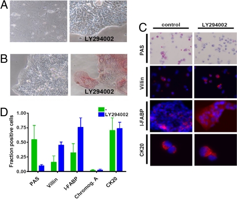
Colon carcinoma is one of the leading causes of death from cancer and is characterized by a heterogenic pool of cells with distinct differentiation patterns. Recently, it was reported that a population of undifferentiated cells from a primary tumor, so-called cancer stem cells (CSC), can reconstitute the original tumor on xenotransplantation. Here, we show that spheroid cultures of these colon CSCs contain expression of CD133, CD166, CD44, CD29, CD24, Lgr5, and nuclear beta-catenin, which have all been suggested to mark the (cancer) stem cell population. More importantly, by using these spheroid cultures or freshly isolated tumor cells from multiple colon carcinomas, we now provide compelling evidence to indicate that the capacity to propagate a tumor with all differentiated progeny resides in a single CSC. Single-cell-cloned CSCs can form an adenocarcinoma on xenotransplantation but do not generate the stroma within these tumors. Moreover, they can self-renew and are capable of multilineage differentiation. Further analysis indicated that the lineage decision is dictated by phosphoinositide 3-kinase (PI3K) signaling in CSCs. These data support the hypothesis that tumor hierarchy can be traced back to a single CSC that contains multilineage differentiation capacity, and provides clues to the regulation of differentiation in colon cancers in vivo.


Conflict of interest statement

The authors declare no conflict of interest.

Figures

Fig. 1.
Fig. 1.
Colon spheroid cultures are heterogeneous for CSC marker expression. (A) Injection of colon cancer spheroid cells generate a tumor that closely resembles the original tumor in morphology, as judged by HE (Upper) and Alcian Blue staining (Lower) of paraffin-embedded sections. (B) Upper shows gradual enrichment for the CD133+ cells in a culture derived from a colon cancer liver metastasis. Lower shows that the marker expression of the CD133+ cells is conserved on expansion in vitro because the profiles for CD24/CD29 and CD44/CD166 expression are overlapping for the direct isolated CD133+ cells and the CD133+ cells in a spheroid culture. (C) Immunofluorescence staining of cytospins of a dissociated colon spheroid culture reveals heterogeneity in nuclear localization of β-catenin and Lgr5 expression. A control for the specificity of the Lgr5 antibody is shown in Right, which contains control transfected or Flag-tagged Lgr5 transfected 293T cells stained for anti-FLAG (red) or Lgr-5 (green). Merge shows both antibodies and indicates a complete overlap in antibody stainings.
Fig. 2.
Fig. 2.
Single-cell derived cultures give rise to differentiated adenocarcinomas on subcutaneous injection into nude mice. (A) Schematic representation of single-cell cloning of CSCs. Unstained single cells were sorted from a colon CSC culture into 96-well plates and after expansion formed single-cell-derived cultures. Twenty of 384 wells that received one single cell showed formation of a sphere. Lower shows formation of adenocarcinoma after subcutaneous injection of SCDCs in immunodeficient mice by using HE and Alcian Blue staining. (B) 384 wells were plated with bulk tumor cells from a T2N0M0 colorectal carcinoma. One hundred fifty-seven wells contained a single cell as judged by microscopy. Spheroids that arose were expanded and injected into immunodeficient mice where they formed an adenocarcinoma (Right) that resembles the original human malignancy (Left) as confirmed by HE and Alcian Blue staining. (C) Schematic representation of the mixing experiment by using CD133+ and CD133 cells. CD133+ GFP-transduced CSCs were single-cell deposited into different amounts (10, 50, 100, and 200) of CD133/GFP cells in 96-well plates and 4 spheres formed that were completely GFP+. Doublets were excluded by using stringent settings on FSC-width. Right shows formation of an adenocarcinoma that is GFP+ as confirmed by HE and Alcian Blue staining and GFP immunohistochemistry. (D) Limiting dilution analysis for different populations within the colon spheroid cultures show diverse clonogenic potential. 1, 2, 4, or 6 cells were deposited by FACS in wells from a 96-well plate and the outgrowth of spheres was monitored. Depicted is the calculated fraction of cells containing sphere initiating capacity. Error bars, 95% confidence interval (*, P < 0.05, **, P < 0.01).
Fig. 3.
Fig. 3.
Functional CSCs can be reisolated from single cell-derived tumors. (A) Xenografts derived from SCDCs show CD133+ cells either detected by FACS analysis (Left) or by immuno-fluorescence staining (Right). (Inset) Higher magnification and confirms membrane localization of CD133. (Scale bar, 200 μm.) (B) SCDC-induced xenografts as generated in Fig. 2B, show a distinct CD133+ fraction, comparable to the primary human malignancy. (C) Spheroid cultures can be isolated from tumors derived from SCDCs, show the same spheroid morphology and phenotype (CK-20 and CD133), and are GFP+ in the case of GFP+ SCDC-derived tumors (Inset). (D) Subcutaneous injection of reisolated spheroid cultures induces a tumor with the same differentiated morphology. HE staining (Left), and Alcian Blue staining (Center) on paraffin sections. Right shows tumor immunofluorescent staining for CD133. (Scale bar, 40 μm.)
Fig. 4.
Fig. 4.
Differentiation of SCDCs reveals multilineage differentiation potential both in vitro and in vivo. (A) Cells from a colon cancer spheroid culture show a gradual decrease in colon CSC marker expression on differentiated on an adherent plate in serum containing medium. Differences are observed in the speed at which the markers are lost. Here, 4 and 8 days of differentiation are shown. CD24 and CD133 show most rapid down regulation, followed by CD44. CD29 and CD166 show very limited change in cell surface expression levels. (B) Undifferentiated spheres were embedded in matrigel and were immediately snap frozen or allowed to differentiate for 10 days and then snap frozen. Sections were stained for CD133 (Upper) and CK20 (Lower). (Scale bars, 40 μm.) (A) Cells differentiated in matrigel show evidence for mucin production as shown here by Alcian Blue stain. (A and C) Data are representative for all clones analyzed (see also Fig. S6). (D) High-magnification microscopy reveals heterogeneous cell morphology in crypt-like structures of SCDCs. Both goblet cell-like (arrows) and enterocyte-like differentiation (arch) can be detected. (E) Staining for markers associated with different cell lineages in the colon epithelium of SCDC-derived xenografts (Upper) or in vitro-differentiated SCDCs (Lower). Villin indicates enterocyte-like differentiation. Alcian Blue and PAS staining reveal mucin production associated with goblet-like cell differentiation. Neuroendocrine differentiation is detected by Chromogranin A staining.
Fig. 5.
Fig. 5.
PI-3 kinase inhibition results in an enterocyte-like differentiation pattern. (A) Cell morphology of a differentiated colon spheroid culture on adherent plate with serum containing medium is changed in the presence of Ly294002. (B) Intestinal alkaline phosphatase (IAP) activity reveals induction of IAP activity in presence of Ly294002. (C) Staining of cytospins of differentiated colon spheroid cultures in the absence (Left) or presence (Right) of Ly294002 for PAS, Villin, and CK20. For I-FABP staining of an adherent culture is shown (for cytospins see Fig. S8C). (D) Quantification of PAS, Villin, I-FABP, CK20, and Chromogranin A positivity in differentiated cells.

References

    1. Dalerba P, Cho RW, Clarke MF. Cancer stem cells: Models and concepts. Annu Rev Med. 2007;58:267–284. - PubMed
    1. Vermeulen L, et al. Cancer stem cells: Old concepts, new insights. Cell Death Differ. 2008;15:947–958. - PubMed
    1. Clarke MF, et al. Cancer stem cells: Perspectives on current status and future directions: AACR workshop on cancer stem cells. Cancer Res. 2006;66:9339–9344. - PubMed
    1. O'Brien CA, Pollett A, Gallinger S, Dick JE. A human colon cancer cell capable of initiating tumour growth in immunodeficient mice. Nature. 2007;445:106–110. - PubMed
    1. Ricci-Vitiani L, et al. Identification and expansion of human colon-cancer-initiating cells. Nature. 2007;445:111–115. - PubMed

Publication types

MeSH terms

Substances