PI3-kinase-dependent activation of apoptotic machinery occurs on commitment of epidermal keratinocytes to terminal differentiation
- PMID: 18766172
- PMCID: PMC2650684
- DOI: 10.1038/cr.2008.281
PI3-kinase-dependent activation of apoptotic machinery occurs on commitment of epidermal keratinocytes to terminal differentiation
Abstract
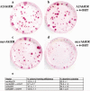
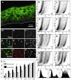
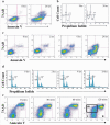
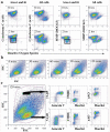
We have investigated the earliest events in commitment of human epidermal keratinocytes to terminal differentiation. Phosphorylated Akt and caspase activation were detected in cells exiting the basal layer of the epidermis. Activation of Akt by retroviral transduction of primary cultures of human keratinocytes resulted in an increase in abortive clones founded by transit amplifying cells, while inhibition of the upstream kinase, PI3-kinase, inhibited suspension-induced terminal differentiation. Caspase inhibition also blocked differentiation, the primary mediator being caspase 8. Caspase activation was initiated by 2 h in suspension, preceding the onset of expression of the terminal differentiation marker involucrin by several hours. Incubation of suspended cells with fibronectin or inhibition of PI3-kinase prevented caspase induction. At 2 h in suspension, keratinocytes that had become committed to terminal differentiation had increased side scatter, were 7-aminoactinomycin D (7-AAD) positive and annexin V negative; they exhibited loss of mitochondrial membrane potential and increased cardiolipin oxidation, but with no increase in reactive oxygen species. These properties indicate that the onset of terminal differentiation, while regulated by PI3-kinase and caspases, is not a classical apoptotic process.
Figures


Similar articles
-
p38 and ERK MAP kinase mediates iron chelator-induced apoptosis and -suppressed differentiation of immortalized and malignant human oral keratinocytes.Life Sci. 2006 Sep 5;79(15):1419-27. doi: 10.1016/j.lfs.2006.04.011. Epub 2006 Apr 26. Life Sci. 2006. PMID: 16697418
-
Caspase activation in the terminal differentiation of human epidermal keratinocytes.Curr Biol. 1999 Apr 8;9(7):361-4. doi: 10.1016/s0960-9822(99)80162-6. Curr Biol. 1999. PMID: 10209121
-
Inhibition of TNF-alpha-induced neutrophil apoptosis by crystals of calcium pyrophosphate dihydrate is mediated by the extracellular signal-regulated kinase and phosphatidylinositol 3-kinase/Akt pathways up-stream of caspase 3.J Immunol. 2000 Nov 15;165(10):5798-806. doi: 10.4049/jimmunol.165.10.5798. J Immunol. 2000. PMID: 11067939
-
Effects of the cyclin-dependent kinase inhibitor CYC202 (R-roscovitine) on the physiology of cultured human keratinocytes.Biochem Pharmacol. 2005 Sep 15;70(6):824-36. doi: 10.1016/j.bcp.2005.06.005. Biochem Pharmacol. 2005. PMID: 16011834
-
Transient activation of FOXN1 in keratinocytes induces a transcriptional programme that promotes terminal differentiation: contrasting roles of FOXN1 and Akt.J Cell Sci. 2004 Aug 15;117(Pt 18):4157-68. doi: 10.1242/jcs.01302. J Cell Sci. 2004. PMID: 15316080
Cited by
-
Neutral evolution of snoRNA Host Gene long non-coding RNA affects cell fate control.EMBO J. 2024 Sep;43(18):4049-4067. doi: 10.1038/s44318-024-00172-8. Epub 2024 Jul 25. EMBO J. 2024. PMID: 39054371 Free PMC article.
-
Caspase activation in tumour-infiltrating lymphocytes is associated with lymph node metastasis in oral squamous cell carcinoma.J Pathol. 2023 Sep;261(1):43-54. doi: 10.1002/path.6145. Epub 2023 Jul 13. J Pathol. 2023. PMID: 37443405 Free PMC article.
-
Biodegradable Gelatin Microcarriers Facilitate Re-Epithelialization of Human Cutaneous Wounds - An In Vitro Study in Human Skin.PLoS One. 2015 Jun 10;10(6):e0128093. doi: 10.1371/journal.pone.0128093. eCollection 2015. PLoS One. 2015. PMID: 26061630 Free PMC article.
-
A protein phosphatase network controls the temporal and spatial dynamics of differentiation commitment in human epidermis.Elife. 2017 Oct 18;6:e27356. doi: 10.7554/eLife.27356. Elife. 2017. PMID: 29043977 Free PMC article.
-
Actin filament dynamics impacts keratinocyte stem cell maintenance.EMBO Mol Med. 2013 Apr;5(4):640-53. doi: 10.1002/emmm.201201839. EMBO Mol Med. 2013. PMID: 23554171 Free PMC article.
References
-
- Jones PH, Simons BD, Watt FM. Sic Transit Gloria. Farewell to the epidermal transit amplifying cell? Cell Stem Cell. 2007;1:371–381. - PubMed
-
- Watt F. Epidermal stem cells. In: Marshak DR, Gottlieb D, editors. Stem Cell Biology. Cold Spring Harbor Laboratory Press; Cold Spring Harbor: 2001. pp. 439–453.
-
- Green H. Terminal differentiation of cultured human epidermal cells. Cell. 1977;11:405–416. - PubMed
-
- Adams J, Watt FM. Fibronectin inhibits the terminal differentiation of human keratinocytes. Nature. 1989;340:307–309. - PubMed
-
- Gandarillas A, Goldsmith LA, Gschmeissner S, Leigh IM, Watt FM. Evidence that apoptosis and terminal differentiation of epidermal keratinocytes are distinct processes. Exp Dermatol. 1999;8:71–79. - PubMed
Publication types
MeSH terms
Substances
Grants and funding
LinkOut - more resources
Full Text Sources
Other Literature Sources
Miscellaneous

