. 2008 Sep;29(3):99-110.
doi: 10.1080/13816810802206499.

Choroideremia: analysis of the retina from a female symptomatic carrier

Affiliations

Choroideremia: analysis of the retina from a female symptomatic carrier

Vera L Bonilha et al. Ophthalmic Genet. 2008 Sep.

Abstract

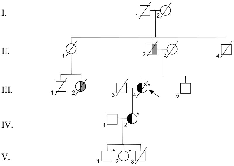
Purpose: To define the retinal pathology in a 91 year-old affected matriarch of a three-generation choroideremia family with multiple manifesting carriers.

Methods: Tissue from three different retinal areas was processed for immunohistochemistry. The macular area was processed for transmission electron microscopy. Cryosections were studied by indirect immunofluorescence, using well-characterized antibodies to cone cytoplasm, rhodopsin and cone opsins. The affected donor eyes were compared to a postmortem matched normal eye.

Results: The retina displayed areas of severe degeneration, with no photoreceptor outer segments, photoreceptor nuclear atrophy, and atrophy of the inner retina. Other retinal areas were near to normal. The RPE was severely degenerated, with thinning, pigment clumping and sub-epithelial debris deposition in all the areas examined. The choroid displayed depigmentation. Labeling with cone opsin antibodies revealed that cones were drastically affected: blue opsin was almost completely absent, while red/green opsins were distributed along the entire plasma membrane of the cell. Rhodopsin was also distributed along the entire rod plasma membrane. Ultrastructural analysis of the affected macula revealed the absence of RPE apical microvilli and basal infoldings. Instead, RPE's basal surface and choroid displayed the presence of banded fibers composed of clumps of wide-spacing collagen. Bruch's membrane was filled with vesicular structures, some smooth and others with bristle-like projections.

Conclusions: The histological data suggests that the clinical manifestation in this donor is related to degenerative changes in the retina, RPE, and choroid.


Figures

Fig. 1
Fig. 1
Pedigree of a family with an X-linked choroideremia showing the symptomatic carrier member (III.4) on whom the postmortem analysis was performed. * Molecularly confirmed to carry 1413+1 G>A in CHM; II-2: By report, totally blind by early 50’s; III-2: By report, legally blind in adulthood; III-4: Propositus. Night blindness & central vision loss late in life (>75yo); IV-2: Night blindness & significant central vision loss in 50’s. Abnormal rod & cone responses on ERG; V-1: Salt and pepper retinopathy; asymptomatic at 34 years old; V-2: Mottling of inferior RPE; asymptomatic at 33 years old. The symbol x indicates patients examined personally; hatched symbols, patients who are affected by history but not personally examined; arrow, proband; single oblique line, deceased.
Fig. 2
Fig. 2
Degeneration in the retina of a female symptomatic carrier of choroideremia. Human 1μm plastic sections of both a matched control and an affected choroideremia stained with toluidine blue. Top panel corner depicts a fundus image of the studied eye with a schematic drawing of the regions cut and processed for cryosectioning (A). Morphology of control retina in the perimacular region of the affected eye (B). (C1, D2, E3) The retina of the affected donor displayed different degrees of photoreceptor degeneration in each of the regions observed. In C1 some pigmented cells are seen invading the degenerating retina (arrowheads). (F1, G2, H3). RPE of the affected donor displayed typical thinning, pigment clumping, basal laminar deposits. In G2 extensive accumulation of amorphous material underneath the RPE layer is observed (*) while in H3 the presence of inflammatory cells (small arrows) is observed in the choroid. (I4, J4, K4) Different areas of the affected donor displaying deposition of drusen under the RPE (large arrows), RPE thinning and pigment clumping, photoreceptor degeneration and the presence of giant lipophilic drops in the choroid (**). RPE= retinal pigment epithelium; Ph=photoreceptors; ONL= outer nuclear layer; OPL= outer plexiform layer; INL= inner nuclear layer; IPL= inner plexiform layer; GCL= ganglion cell layer. Bar= 200μm.
Fig. 3
Fig. 3
Cone cytoplasmic marker distribution is normal in a female symptomatic carrier of choroideremia. Human paraffin sections of both a matched control (A) and affected choroideremia donor (B, C, D) were labeled with an antibody specific to cone cytoplasm (green) while cell nuclei were labeled with TO-PRO-3 (blue). Sections were analyzed using a Leica laser scanning confocal microscope. Comparison of the samples showed that there was no significant difference in the overall morphology of the control and affected donor cones. Bar = 40μm.
Fig. 4
Fig. 4
Red/Green cone opsins are distributed along the entire plasma membrane of this cone type in a female symptomatic carrier of choroideremia. Human paraffin sections of both a matched control (A) and affected choroideremia donor (B, C, D) were labeled with antibodies specific to red/green cone opsins (green) while cell nuclei were labeled with TO-PRO-3 (blue). Sections were analyzed using a Leica laser scanning confocal. Comparison of the samples showed that red/green cone opsins are distributed along the entire plasma membrane of this cone type, from the tip of the outer segment to the synaptic base in all the observed regions of the affected eye. Bar = 40μm.
Fig. 5
Fig. 5
Blue cone opsins are significantly decreased in this cone type in a female symptomatic carrier of choroideremia. Human paraffin sections of both a matched control (A) and affected choroideremia donor (B, C, D) were labeled with antibodies specific to blue cone opsins (green) while cell nuclei were labeled with TO-PRO-3 (blue). Sections were analyzed using a Leica laser scanning confocal microscope. Comparison of the samples showed that blue cone opsins are significantly decreased in all the observed regions of the affected eye. Region 2 displays a few cones that still express blue opsin but it is distributed throughout the inner and outer segments of the cones (arrow). Bar = 40 μm.
Fig. 6
Fig. 6
Rhodopsin is distributed along the entire plasma membrane of the rods in a female symptomatic carrier of choroideremia. Human paraffin sections of both a matched control (A) and affected choroideremia donor (B, C, D) were labeled with antibodies specific to rhodopsin (Alexa488, green) while cell nuclei were labeled with TO-PRO-3 (blue). Sections were analyzed using a Leica laser scanning confocal microscope. Comparison of the samples showed that rhodopsin is distributed along the entire plasma membrane of rods, from the tip of the outer segment to the synaptic base in all the observed regions of the affected eye. Bar = 40μm.
Fig. 7
Fig. 7
Ultrastructural evidence of RPE degeneration in a female symptomatic carrier of choroideremia. The ultrastructure of RPE and Bruch’s membrane was analyzed by TEM. Observation at low magnification showed photoreceptor outer segments (POS) lying on top of a collapsed RPE apical surface while a control-matched eye displayed several apical microvilli supporting the POS (A). The matched control displayed extensive basal infoldings (BI) in their basal surface on top of a well-structured aged Bruch’s membrane (BM). The choroideremia donor cytoplasm was filled with small vacuoles and pigments (P) (C). Observation of RPE’s basal surface in the choroideremia donor revealed absence of basal infoldings and the presence of banded fibers composed of clumps of wide-spacing collagen (arrows, D, E, F). The choroid (Ch), Bruch’s membrane (BM) and the space between the RPE basal membrane was filled with vesicular structures, some smooth and others like bristle-coated vesicles (D, E). Higher magnification of this area is shown in E. Another area of the choroideremia donor eye displayed drusen (Dr) and beneath it Bruch’s membrane contained residual bodies, vesicular material and filaments (F). Bars: C, D, F = 2μm and A, B, E =1μm.

References

    1. Lyon M. Gene action in X chromosome of mouse (Mus musculus L.) Nature. 1961;190:372–373. - PubMed
    1. Endo K, Yuzawa M, Ohba N. Choroideremia associated with subretinal neovascular membrane. Acta Ophthalmol Scand. 2000;78:483–6. - PubMed
    1. Potter MJ, Wong E, Szabo SM, McTaggart KE. Clinical findings in a carrier of a new mutation in the choroideremia gene. Ophthalmology. 2004;111:1905–1909. - PubMed
    1. Rubin ML, Fishman RS, McKay RA. Choroideremia: study of a family and literature review. Arch Ophthalmol. 1966;76:563–574. - PubMed
    1. Mura M, Sereda C, Jablonski MM, MacDonald IM, Iannaccone A. Clinical and functional findings in choroideremia due to complete deletion of the CHM gene. Arch Ophthalmol. 2007;125:1107–1113. - PubMed

Publication types

MeSH terms

LinkOut - more resources