. 2008 Nov 1;105(4):1027-37.
doi: 10.1002/jcb.21902.

E-cadherin dis-engagement activates the Rap1 GTPase

Affiliations

E-cadherin dis-engagement activates the Rap1 GTPase

Sirisha Asuri et al. J Cell Biochem. .

Abstract

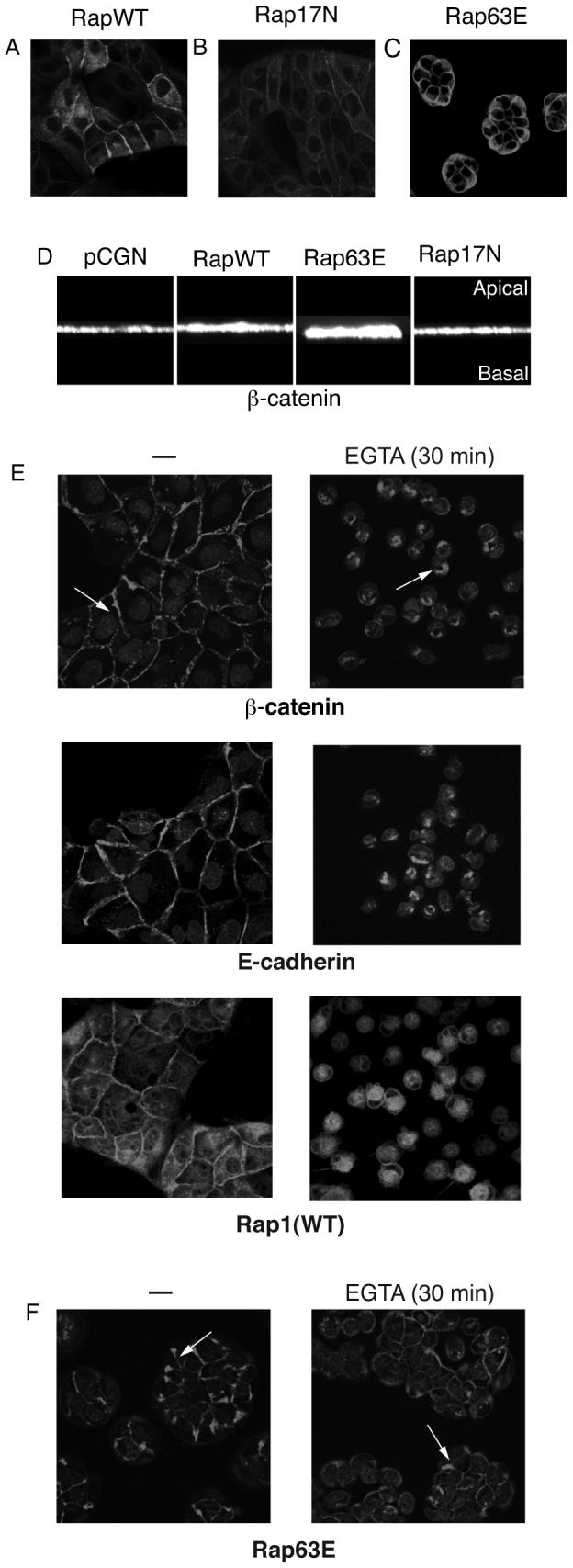
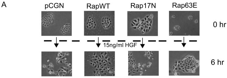
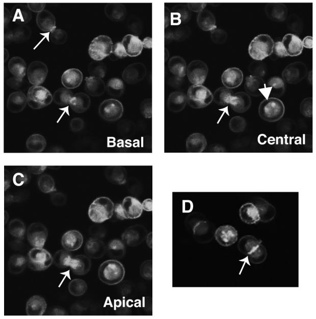
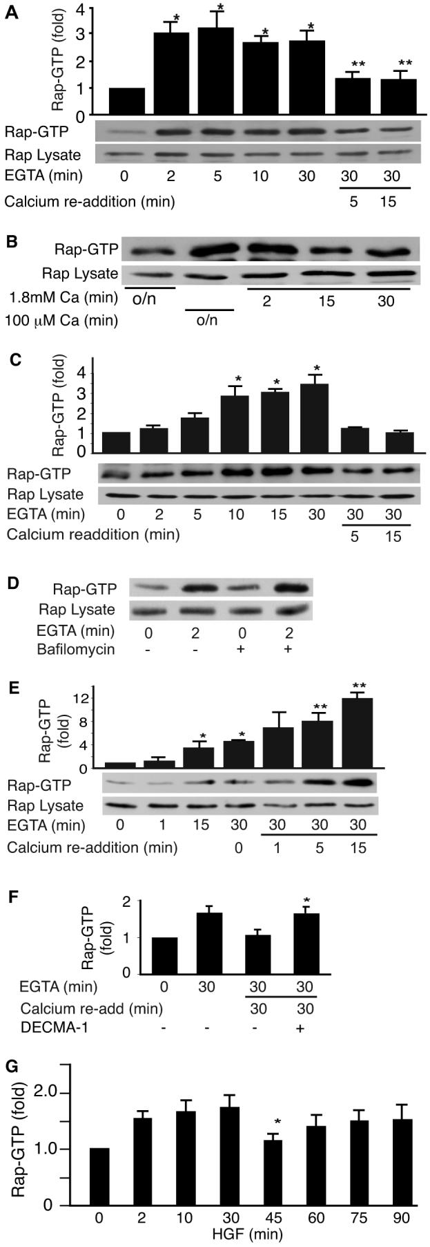
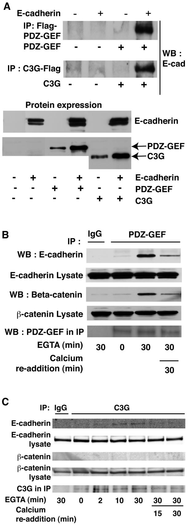
E-cadherin based adherens junctions are finely regulated by multiple cellular signaling events. Here we show that the Ras-related Rap1 GTPase is enriched in regions of nascent cell-cell contacts and strengthens E-cadherin junctions: constitutively active Rap1 expressing MDCK cells exhibit increased junctional contact and resisted calcium depletion-induced cell-cell junction disruption. E-cadherin disengagement activated Rap1 and this correlated with E-cadherin association with the Rap GEFs, C3G and PDZ-GEF I. PDZ-GEF I associated with E-cadherin and beta-catenin whereas C3G interaction with E-cadherin did not involve beta-catenin. Knockdown of PDZ-GEF I in MDCK cells decreased Rap1 activity following E-cadherin junction disruption. We hereby show that Rap1 plays a role in the maintenance and repair of E-cadherin junctions and is activated via an "outside-in" signaling pathway initiated by E-cadherin and mediated at least in part by PDZ-GEF I.


Figures

Fig. 1
Fig. 1. Rap1 localizes to regions of cell–cell contact in epithelial cells
MDCK cells stably expressing HA-tagged wild type Rap1A, Rap1A-WT (A), dominant negative Rap1A-17N (B) or constitutively active Rap1A-63E (C) were stained using HA antibody and imaged using confocal microscopy. D: The height of adherens junctions was determined by detection of β-catenin. The images were analyzed using the VOX program (VayTek Inc., Fairfield, IA) to obtain a 90° rotation and vertical perception of β-catenin localization in three-dimensional space. E: Rap1A-WT expressing MDCK cells were subject to calcium switch and β-catenin, E-cadherin and HA-tagged Rap1A detected by immunofluorescence microscopy. Following extracellular calcium chelation, adherens junctions were disrupted and proteins endocytosed. Whereas the Rap1A-WT cells underwent complete dissolution of adherens junctions and endocytosis of β-catenin (arrow) Rap1A-63E cells retained most cell–cell contacts and β-catenin on their membrane (F). Note also the increased β-catenin staining at cell–cell contacts in Rap1A-63E expressing colonies before treatment with EGTA. Data representative of three independent experiments.
Fig. 2
Fig. 2. Constitutively active Rap1 strengthens E-cadherin junctions
A: MDCK cells stably transfected with empty pCGN vector, or vector encoding wild type Rap1A (RapWT), constitutively active Rap1A mutant (Rap63E) or dominant negative Rap1 mutant (Rap17N) were treated with HGF for 6 h. Cells transfected with pCGN, pCGN Rap1A-WT or Rap1A-17N underwent dissolution of cell–cell junctions followed by cell scattering whereas Rap1A-63E expressing cells did not undergo junction disruption. All images were obtained at the same magnification. Representative of at least three independent experiments.
Fig. 3
Fig. 3. Rap1 localizes to nascent cell–cell contacts during cell junction formation
MDCK cells stably expressing GFP-Rap1A-WT were trypsinized, vigorously pipetted to disrupt cell–cell junctions, and re-plated. Localization of GFP-Rap1A in these cells was then followed using live cell confocal microscopy. A–C: Images collected on the same microscopic field of the plate from near the basal (A) to near the apical (C) surface of the cells. GFP-Rap1A was mainly intracellular with a ring of Rap1 on the plasma membrane (down facing arrows) in single cells whereas GFP-Rap1A is highly enriched when cells make contact and begin to establish cell junctions (upward facing arrow). D: Field showing two cells establishing a nascent cell–cell contact and GFP-Rap1A enriched at the cell junction. Data representative of three or more independent experiments.
Fig. 4
Fig. 4. Rap1 is activated during disruption of E-cadherin based adherens junctions
A: Calcium switch induced activation of Rap1. MDCK cell junctions were disrupted by addition of 4 mM EGTA to the medium to chelate calcium and reformed by adding back 4 mM CaCl2 (calcium switch). Data representative of at least 10 independent experiments. A representative blot and pooled data from 5 to 9 experiments is shown, mean +/− SEM, P<0.01 versus t=0 (*) or t=30 (**) min. B: Rap1 activation during low calcium switch. Rap1 was activated following overnight incubation of MDCK cells in low calcium (100 μM) medium (lane 2). This activation was reversed when cells were returned to regular calcium medium for 2, 15, or 30 min (lanes 3–5). C: Rap1 was activated by Ca2+ removal in Fisher rat thyroid (FRT) epithelial cells. P<0.05 versus t=0, n=3. D: 30 min pretreatment with 100 nM bafilomycin did not prevent calcium chelation from rapidly activating Rap1. Representative of three separate experiments. E: Rap1 activation following calcium switch in HUVECs. A minimal activation of Rap1 during the disruption of adherens junctions was seen in endothelial cells (lanes 3 and 4 P<0.05) with a stronger activation of Rap1 during re-addition of 1.8 mM CaCl2 (lanes 6–8, P<0.05 vs. t=30 min). F: Incubation of cells with E-cadherin antibody, DECMA-1 to prevent homotypic binding prevented Rap1 deactivation. P<0.05 versus goat IgG control, n=3. G: HGF induced activation of Rap1. MDCK cells were treated with HGF (15 ng/ml) for the specified times and Rap1GTP levels measured. Graph shows biphasic increase in RapGTP following treatment with HGF (mean +/− SEM) from four independent experiments. P<0.025 versus t=30 min.
Fig. 5
Fig. 5. E-cadherin co-precipitates with PDZ-GEF I and C3G
A: E-cadherin was exogenously expressed in 293T cells along with either Flag tagged PDZ-GEF I or C3G. Flag antibody was used to precipitate GEFs prior to blotting for E-cadherin. E-cadherin co-precipitated with both PDZ-GEF I (top panel) and C3G (middle panel). Expression of proteins is shown below. B: Endogenous E-cadherin and β-catenin associate with PDZ-GEF I in MDCK cells. MDCK cells were subject to calcium switch and PDZ-GEF antibody used to immunoprecipitate the endogenous GEF. Association of endogenous E-cadherin with PDZ-GEF was greatly enhanced upon disruption of junctions with EGTA for 30 min but returned to near basal levels following Ca2+ re-addition. β-Catenin also coprecipitated with the E-cadherin and PDZ-GEF I complex (lower panels). C: E-cadherin but not β-catenin co-immunoprecipitated with C3G in MDCK cells. MDCK cells were calcium switched as above and C3G antibody used for immunoprecipitation. Data representative of at least three independent experiments.
Fig. 6
Fig. 6. Knockdown of MDCK cell PDZ-GEF I reduces the ability of cell junction disruption to activate Rap1
A: Transfection of two different PDZGEF siRNAs resulted in decreased expression of the GEF 48 hr later. B: MDCK cells were transfected with control or two different PDZ-GEF I siRNAs (75 mM siRNA#3 shown). Forty-eight hours later calcium switch was performed as indicated and RapGTP levels measured using the RalGDS-RBD pull down. Glyceraldehyde 3 phosphate dehydrogenease and total Rap1 expression levels indicate equal loading. C: Graph shows quantitation of the data represented in (B) (mean +/− SEM from three separate experiments using siRNA #3).

Similar articles

Cited by

References

    1. Adams CL, Chen YT, Smith SJ, Nelson WJ. Mechanisms of epithelial cell-cell adhesion and cell compaction revealed by high-resolution tracking of E-cadherin-green fluorescent protein. J Cell Biol. 1998;142:1105–19. - PMC - PubMed
    1. Adams CL, Nelson WJ, Smith SJ. Quantitative analysis of cadherin-catenin-actin reorganization during development of cell-cell adhesion. J Cell Biol. 1996;135:1899–911. - PMC - PubMed
    1. Arthur WT, Noren NK, Burridge K. Regulation of Rho family GTPases by cell-cell and cell-matrix adhesion. Biol Res. 2002;35:239–46. - PubMed
    1. Arthur WT, Quilliam LA, Cooper JA. Rap1 promotes cell spreading by localizing Rac guanine nucleotide exchange factors. J Cell Biol. 2004;167:111–22. - PMC - PubMed
    1. Balzac F, Avolio M, Degani S, Kaverina I, Torti M, Silengo L, Small JV, Retta SF. E-cadherin endocytosis regulates the activity of Rap1: a traffic light GTPase at the crossroads between cadherin and integrin function. J Cell Sci. 2005;118:4765–83. - PubMed

Publication types

LinkOut - more resources