. 2008 Oct 1;130(39):13132-9.
doi: 10.1021/ja8045469. Epub 2008 Sep 4.

Ultrafast dynamics of flavins in five redox states

Affiliations

Ultrafast dynamics of flavins in five redox states

Ya-Ting Kao et al. J Am Chem Soc. .

Abstract

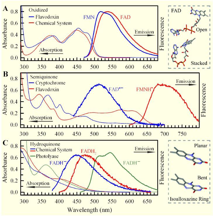
We report here our systematic studies of excited-state dynamics of two common flavin molecules, FMN and FAD, in five redox states--oxidized form, neutral and anionic semiquinones, and neutral and anionic fully reduced hydroquinones--in solution and in inert protein environments with femtosecond resolution. Using protein environments, we were able to stabilize two semiquinone radicals and thus observed their weak emission spectra. Significantly, we observed a strong correlation between their excited-state dynamics and the planarity of their flavin isoalloxazine ring. For a bent ring structure, we observed ultrafast dynamics from a few to hundreds of picoseconds and strong excitation-wavelength dependence of emission spectra, indicating deactivation during relaxation. A butterfly bending motion is invoked to get access to conical intersection(s) to facilitate deactivation. These states include the anionic semiquinone radical and fully reduced neutral and anionic hydroquinones in solution. In a planar configuration, flavins have a long lifetime of nanoseconds, except for the stacked conformation of FAD, where intramolecular electron transfer between the ring and the adenine moiety in 5-9 ps as well as subsequent charge recombination in 30-40 ps were observed. These observed distinct dynamics, controlled by the flavin ring flexibility, are fundamental to flavoenzyme's functions, as observed in photolyase with a planar structure to lengthen the lifetime to maximize DNA repair efficiency and in insect type 1 cryptochrome with a flexible structure to vary the excited-state deactivation to modulate the functional channel.


Figures

Figure 1
Figure 1
Steady-state absorption and emission spectra of flavins in five redox states. (A) oxidized state, (B) radical state, and (C) fully-reduced state. The excitation wavelengths are 400 nm for oxidized FAD and FMN, 420 nm for anionic radical FAD•-, 580 nm for neutral radical FMNH, and 360 nm for fully-reduced FADH- and FADH2. The fluorescence emissions of FAD•- in the cryptochrome and FADH- and FADH2 in solution are very weak. Upper right frame: Schematic representation of FAD in open and stacked conformations. The stacked conformation was adopted from U—shaped FAD in photolyase. Lower right frame: Schematic representation of the isoalloxazine ring in planar and “butterfly” bent conformations.
Figure 2
Figure 2
Femtosecond-resolved oxidized flavin dynamics in solution and in flavodoxin. (A) Normalized fluorescence transients of excited FAD gated at 490 and 530 nm with removal of the long lifetime components in nanosecond. The complete signal is shown in inset with comparison to the transient of FMN* in the flavodoxin mutant with a predominant lifetime in nanosecond. (B) Transient-absorption detection of oxidized FAD in solution for various pump/probe schemes. Note that the nanosecond decay from non-ET dynamics has been subtracted for clarity. The coupled solvation and ET dynamics is in 4-5 ps and the charge recombination takes 30-40 ps.
Figure 3
Figure 3
Femtosecond-resolved fluorescence transients of anionic radical FAD•- in the insect cryptochrome (ApCRY1) gated at 550 nm (A) and of neutral radical FMNH in the flavodoxin mutant gated at 680 nm (B). Insets show the excitation-wavelength dependence of weak FAD•- emission spectra, indicating deactivation during relaxation, and the excitation-wavelength independence of weak FMNH emission spectra, resulting from the lowest excited-state emission. For all emission spectra, the Raman scattering signals at the blue side of emission peaks were all removed for clarity.
Figure 4
Figure 4
Femtosecond-resolved fluorescence transients of fully-reduced flavins. Fully-reduced anionic FADH- and FMNH- in the proteins (A), photolyase and the flavodoxin mutant, and in solution (B). (C) Fully-reduced neutral FADH2 in solution. Insets show strong excitation-wavelength dependence of weak FADH- and FADH2 emission spectra, indicating deactivation during relaxation. The Raman scattering signals at the blue side of emission peaks were all removed for clarity.
Figure 5
Figure 5
Femtosecond-resolved transient-absorption detection of fully-reduced flavins in solution upon 325-nm excitation with probing from 690 to 585 nm. (A) FMNH- (B) FADH- and (C) FADH2. The dynamics of each species at different probing wavelengths are all similar.
Figure 6
Figure 6
Schematic potential energy landscape of fully-reduced flavins in solution and in proteins. With a flexible bending motion to access CI(s), the flavin molecule in solution exhibits complex deactivation dynamics. In photolyase and the flavodoxin mutant, the bending motion is restricted at the local region and the flavin molecule could not get access to CI(s), leading to a long nanosecond lifetime. The bending coordinate ( θ) could be defined for the “butterfly” motion of the isoalloxazine ring along N5—N10 axis.
Figure 7
Figure 7
Comparative studies on fully-reduced flavins with various excitation wavelengths, solvent viscosity, and temperatures. (A) Normalized fluorescence transients of excited FADH- in solution with 400 and 325 nm excitation. (B) Normalized absorption transients of excited FMNH- in solution with 400 and 325 nm excitation and 585 nm probing. (C) Normalized absorption transients of excited FMNH- in 0% and 50% glycerol solutions. (D) Normalized fluorescence transients of excited FADH- in photolyase with 10%, 30% and 50% glycerol buffers. (E) Normalized fluorescence transients of FADH- in photolyase under 0°C, 12 °C and 25°C.
Scheme1
Scheme1
Structure formulae of flavin in oxidized state.
Scheme 2
Scheme 2
Different redox states of flavin (FAD) under physiological conditions

References

    1. Walsh C. Acc. Chem. Res. 1980;13:148–155.
    1. Massey V, Hemmerich P. Biochem. Soc. Trans. 1980;8:246–257. - PubMed
    1. Müller F. The Flavin Redox-System and Its Biological Function. In: Boschke FL, editor. Topics in Current Chemistry. Vol. 108. Springer -Verlag; Berlin: 1983. pp. 71–107. - PubMed
    1. Müller F, editor. Chemistry and Biochemistry of Flavoenzymes. I-III. CRC Press; Boca Raton, FL: 19901991.
    1. Fraaije MW, Mattevi A. Trends Biochem. Sci. 2000;25:126–132. - PubMed

Publication types