. 2008 Oct;8(10):3461-7.
doi: 10.1021/nl802351p. Epub 2008 Sep 4.

Photothermal optical coherence tomography of epidermal growth factor receptor in live cells using immunotargeted gold nanospheres

Affiliations

Photothermal optical coherence tomography of epidermal growth factor receptor in live cells using immunotargeted gold nanospheres

Melissa C Skala et al. Nano Lett. 2008 Oct.

Abstract

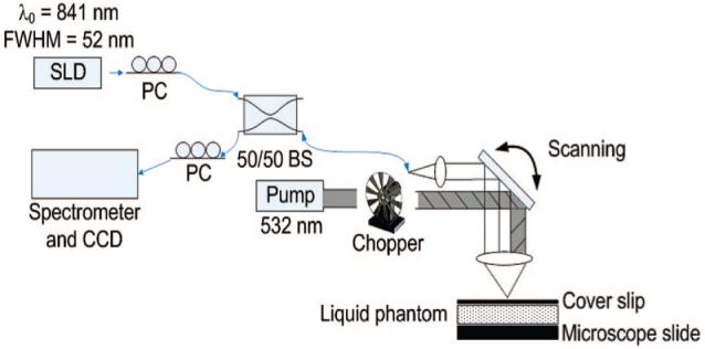
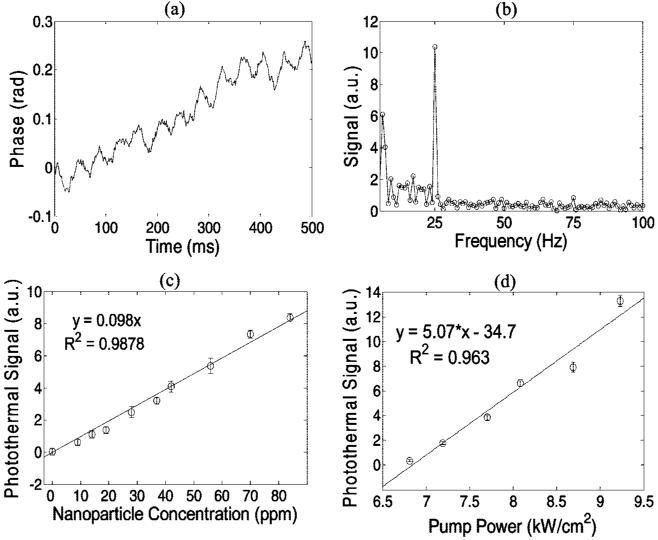
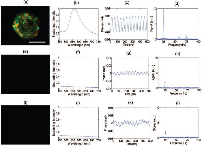
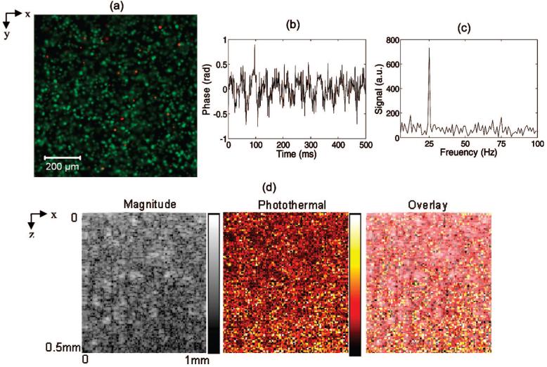
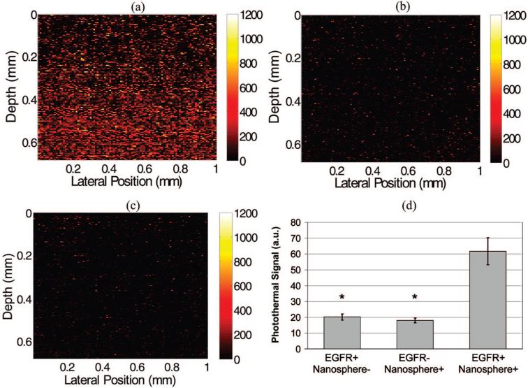
Molecular imaging is a powerful tool for investigating disease processes and potential therapies in both in vivo and in vitro systems. However, high resolution molecular imaging has been limited to relatively shallow penetration depths that can be accessed with microscopy. Optical coherence tomography (OCT) is an optical analogue to ultrasound with relatively good penetration depth (1-2 mm) and resolution (approximately 1-10 microm). We have developed and characterized photothermal OCT as a molecular contrast mechanism that allows for high resolution molecular imaging at deeper penetration depths than microscopy. Our photothermal system consists of an amplitude-modulated heating beam that spatially overlaps with the focused spot of the sample arm of a spectral-domain OCT microscope. Validation experiments in tissuelike phantoms containing gold nanospheres that absorb at 532 nm revealed a sensitivity of 14 ppm nanospheres (weight/weight) in a tissuelike environment. The nanospheres were then conjugated to anti-EGFR, and molecular targeting was confirmed in cells that overexpress EGFR (MDA-MB-468) and cells that express low levels of EGFR (MDA-MB-435). Molecular imaging in three-dimensional tissue constructs was confirmed with a significantly lower photothermal signal (p<0.0001) from the constructs composed of cells that express low levels of EGFR compared to the overexpressing cell constructs (300% signal increase). This technique could potentially augment confocal and multiphoton microscopy as a method for deep-tissue, depth-resolved molecular imaging with relatively high resolution and target sensitivity, without photobleaching or cytotoxicity.


Figures

Figure 1
Figure 1
Schematic of the photothermal OCT setup. PC, polarization controller; SLD, super luminescent diode; BS, beam splitter.
Figure 2
Figure 2
Phase of the tissuelike phantom (polystyrene spheres with μs = 100 cm-1) with 84 ppm nanospheres and 25 Hz pump frequency (a). The definition of the photothermal signal (b) in the Fourier transformed phase is the peak at 25 Hz minus the background (27-50 Hz). The photothermal signal is linearly dependent on the nanoparticle concentration (c) in the scattering phantom with a pump beam intensity of 8 kW/cm2, and the sensitivity of the system is 14 ppm (0 versus 14 ppm, p = 0.006). The photothermal signal is linearly dependent on the pump laser intensity (d) in the tissuelike phantom with 70 ppm nanospheres. All error bars are standard error of N = 10 measurements.
Figure 3
Figure 3
EGFR expression and nanoparticle labeling was confirmed in EGFR+/nanosphere+ cells (a-d), with controls EGFR-/nanosphere+ (e-h) and EGFR+/nanosphere-(i-l) with dark field microscopy (panels a, e, i) and microspectroscopy (panels b, f, j). The phase as a function of time in the photothermal system is plotted for the experimental (panel c) and control groups (panels g, k) and the Fourier Transform of the phase confirms oscillations at 25 Hz (the pump laser modulation frequency) (panels d, h, l). In three repeated experiments, the photothermal signal from overexpressing cell monolayers was at least 9 dB higher than the highest signal from the cells that express low levels of EGFR. The repeated experiments were performed at pump powers of 7.5-8.5 kW/cm2. Scale bar is 10 μm.
Figure 4
Figure 4
A confocal image of a three-dimensional construct in the en face orientation (images of the x-y plane) acquired after applying a live (green) dead (red) fluorescence stain (a) indicates good cell viability and relatively homogeneous cell distribution. Representative photothermal OCT data from a three-dimensional cell construct containing MDA-MB-468 cells (EGFR+) labeled with antibody-conjugated nanospheres (nanosphere+). The phase as a function of time (b) from a representative pixel shows the characteristic 25 Hz frequency component (c). The OCT intensity image, photothermal image, and overlay of intensity/photothermal images of the three-dimensional construct are also shown (d) in the transverse (x-z) plane. Pump power 8.5 kW/cm2.
Figure 5
Figure 5
Images of EGFR expression in three-dimensional cell constructs containing EGFR+ cells (MDA-MB-468) with and without antibody-conjugated nanospheres (a and c, respectively) and EGFR-cells (MDA-MB-435) with antibody-conjugated nanospheres (b). There was a significant increase in the photothermal signal from EGFR overexpressing cell constructs labeled with antibody conjugated nanospheres (d) compared to the two controls (EGFR+/Nanosphere- and EGFR-/Nanosphere+). N = 17 images for each group, (*, p < 0.0001). Pump power 8.5 kW/cm2.

References

    1. Applegate BE, Izatt JA. Opt. Exp. 2006;14(20):9142–9155. - PubMed
    1. Xu C, Ye J, Marks DL, Boppart SA. Opt. Lett. 2004;29(14):1647–1649. - PubMed
    1. Vinegoni C, Bredfeldt J, Marks D, Boppart S. Opt. Express. 2004;12(2):331–341. - PubMed
    1. Yang C, Choma MA, Lamb LE, Simon JD, Izatt JA. Opt. Lett. 2004;29(12):1396–1398. - PubMed
    1. Oldenburg AL, Gunther JR, Boppart SA. Opt. Lett. 2005;30(7):747–9. - PubMed

Publication types