. 2008 Sep 2;6(9):e209.
doi: 10.1371/journal.pbio.0060209.

Redundant mechanisms recruit actin into the contractile ring in silkworm spermatocytes

Affiliations

Redundant mechanisms recruit actin into the contractile ring in silkworm spermatocytes

Wei Chen et al. PLoS Biol. .

Abstract

Cytokinesis is powered by the contraction of actomyosin filaments within the newly assembled contractile ring. Microtubules are a spindle component that is essential for the induction of cytokinesis. This induction could use central spindle and/or astral microtubules to stimulate cortical contraction around the spindle equator (equatorial stimulation). Alternatively, or in addition, induction could rely on astral microtubules to relax the polar cortex (polar relaxation). To investigate the relationship between microtubules, cortical stiffness, and contractile ring assembly, we used different configurations of microtubules to manipulate the distribution of actin in living silkworm spermatocytes. Mechanically repositioned, noninterdigitating microtubules can induce redistribution of actin at any region of the cortex by locally excluding cortical actin filaments. This cortical flow of actin promotes regional relaxation while increasing tension elsewhere (normally at the equatorial cortex). In contrast, repositioned interdigitating microtubule bundles use a novel mechanism to induce local stimulation of contractility anywhere within the cortex; at the antiparallel plus ends of central spindle microtubules, actin aggregates are rapidly assembled de novo and transported laterally to the equatorial cortex. Relaxation depends on microtubule dynamics but not on RhoA activity, whereas stimulation depends on RhoA activity but is largely independent of microtubule dynamics. We conclude that polar relaxation and equatorial stimulation mechanisms redundantly supply actin for contractile ring assembly, thus increasing the fidelity of cleavage.


Conflict of interest statement

Competing interests. The authors have declared that no competing interests exist.

Figures

Figure 1
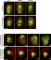
Figure 1. Cytokinesis of Silkworm Spermatocytes
(A) Polarization micrograph of a dividing spermatocyte. (B) Confocal micrographs of a spermatocyte microinjected with rhodamine tubulin (microtubules false colored green), and low-level Alexa Fluor 488 phalloidin (actin false colored red). Actin aggregates appeared at the equatorial microtubule plus ends during early anaphase (0–20), then fused into a contractile ring that bisected the cell (4–45). (C) Astral flagella, as well as naturally, asymmetrically positioned asters, were visible in dividing spermatocytes. Astral flagella were visible only in fixed, immunostained cells (a–c). Astral microtubules were prominent in fixed anaphase spermatocytes (e.g., b). One aster was loosely attached to the spindle (arrow). Occasionally, the asters (arrows) in fixed (a and c) and live (d and e) spermatocytes (arrows) were naturally positioned on the same side of the spindle. a and d, metaphase; b, c and e. anaphase. Fixed cells were double-immunolabeled for actin and tubulin; live cells were labeled as in (B). Time in min. Time 0 in (A and B), anaphase onset. Bars, 10 μm.
Figure 2
Figure 2. Cortical Actin Filaments Were Excluded from the Polar Cortex Bordering the Asymmetrically Distributed Asters
(A) Both asters (arrows) were naturally positioned at the upper pole of the spindle. Cortical actin filaments flowed towards the lower polar cortex during early anaphase. Cells in Figure 2 were labeled as in Figure 1B. Time 0 depicts anaphase onset. (B) Cortical actin filaments, excluded by asymmetrically distributed asters (arrows), assembled a contractile ring around the equator of the naturally shifted spindle. Actin aggregates were also present by later anaphase. Time in min. Time 0, anaphase onset. Bars, 10 μm.
Figure 3
Figure 3. Cortical Flow of Actin Filaments Was Driven by Spindle Microtubules
(A) During anaphase (0), when the spindle apparatus was collapsed, and pushed with a microneedle to an arbitrary region of the cell cortex (3), actin filaments flowed to the opposite side of the cell (3–25). This redistribution of actin filaments by spindle microtubules resulted in asymmetric cell division (71). (B) Repositioning and reorganization of the spindle (2) during telophase resulted in a similar scenario, preceded by regression of the original furrow. (C) An anaphase cell (with only one aster visible in this focal plane; 0) was mechanically remodeled by moving spindle microtubules to the upper portion of the cell and collapsing the spindle (shown at top of cell, 5–21). The collapsed spindle was manipulated to detach and relocate first one of its asters (dot in center of cell, 5) and then its second aster (to left of first aster; both asters marked by arrows, 12–21). Possibly due to actin exclusion (5–21) by microtubules from both structures, the contractile ring formed between the collapsed spindle and the pair of detached asters (21–66). This is shown in the schematic diagram (representing the cell in 21). (D) A schematic of the set-up, with fluorescence images of microtubule-driven actin flow blocked by a microneedle. The schematic shows the cell's outline, the location of the needle (gray), and the direction of flow of cortical actin (red), as induced by the collapsed, repositioned spindle (green, at top). The manipulation needle (arrows), which indented but did not pierce the plasma membrane, locally intercepted the actin flow; brighter fluorescence accumulated on the side of the needle that faced the repositioned spindle (1.7–4.5). The three large red objects (3.4–4.5, upper right) are cell division scars, i.e., remnants from former cytokinetic rings. These scar structures can also move around within the cortex during cortical flow. Note: despite removal of the holding needle, the spindle is not depicted as a giant aster, since the reorganization from monopolar spindle to aster takes at least several minutes. Cells in (A–D) were labeled as in Figure 1B. Time in min. Time 0 in (A), (C), and (D), mid to late anaphase; in (B), telophase. Bars, 10 μm.
Figure 4
Figure 4. Cortical Stiffness Correlates with Local Density of Cortical Actin
(A) After the spindle was repositioned and collapsed to induce cortical flow, we tested the stiffness of the actin-dense region of the cortex. (a) The needle (marked with asterisks) in its initial position, tangential to the actin-rich cortex. (b) After the needle was pushed against the cortex, the stiff cortex deflected the needle. (B) After rotating the cell in (A) by 180°, the cortical stiffness of the actin-depleted region of the cortex was tested. (a) The needle (marked with asterisks) in its initial position, tangential to actin-depleted cortex. (b) After the needle was pushed against the cortex, the relaxed cortex was deformed by the needle without deflecting it. F-actin was labeled using low-level Alexa Fluor 488 phalloidin to monitor flow. Bars, 10 μm.
Figure 5
Figure 5. Interdigitating Microtubule Plus Ends Hosted De Novo Actin Assembly and Delivered Actin Aggregates to Equator
(A) Emergence (0–2) and growth (2–10) of nascent actin fluorescence at the equatorial microtubule plus-end overlap region reflect de novo assembly of actin aggregates. (B) A splaying microtubule bundle (arrowheads) delivered the actin aggregate into the ingressing furrow. Cells in (A) and (B) were labeled as in Figure 1B. (C) De novo assembly of actin aggregates was not inhibited by microtubule stabilization (compare to (A) and Figure 1B). Microtubules were labeled and stabilized by Oregon green paclitaxel, and actin filaments were labeled by rhodamine phalloidin. Insets: actin channel. Time in min. Time 0 in (A and C), immediately before anaphase onset; in (B), late anaphase. Bars, 10 μm.
Figure 6
Figure 6. Redistribution of Cortical Actin, but Not Equatorial Stimulation, Was Dependent on Microtubule Dynamics
(A) A schematic showing remodeling of the central spindle by micromanipulation. A post-anaphase spindle (a, green) was collapsed using a micromanipulation needle (b, gray). The manipulation created two lateral microtubule bundles, with the exposed microtubule plus ends of the bundles pointing toward opposite sides of the cortex. The interior ends of the bundles flanked chromosomes (c, blue), kinetochores (c, purple) and centrosomes (c, orange). If the collapsed spindle was brought close to the cortex and held in place by a microneedle, it reorganized into a monopolar spindle (d). If the holding needle was removed, the spindle usually reorganized into a giant aster (e). (B) Actin exclusion was inhibited when the remodeled spindle was stabilized by paclitaxel. Neither the stabilized lateral spindle (8) nor the evolving monopolar spindle (12–22) induced unidirectional flow of actin filaments (compare to Figure 3). Cells in (B–F) were labeled as in Figure 5C. (C) Actin aggregates were assembled at the plus ends of stabilized, laterally oriented microtubule bundles (0–2, arrowheads), and were delivered to the non-equatorial cortex where splaying microtubule bundles were in contact with the cortex (2–12, arrows). (D) A monopolar spindle (2) gradually splayed its microtubule bundles toward the cell cortex (2–21), which delivered actin aggregates (2, arrowheads) and induced a furrow (11–21, arrows). The furrow eventually regressed (61). (E) Tracking of an actin aggregate delivered from the plus ends of one microtubule bundle to the cell cortex. The giant aster had chromosomes in its center and microtubule plus ends pointing outward, as in (A, panel e). When the aster was placed near the cell cortex using a microneedle, an actin aggregate moved away from the microtubule bundle plus ends and merged into the cortex (box, 0–8.8). (F) The region of interest in (E) is shown with additional intervening time points to highlight the increasing cortical fluorescence caused by merging of actin aggregates. Time in min. Time 0 in (B), (D), and (E), mid to late anaphase; in (C), telophase; in (F), identical to 0 in (E). Bars, 10 μm.
Figure 7
Figure 7. Equatorial Stimulation, but Not Redistribution of Cortical Actin, Was Dependent on RhoA Activity
(A) A cytokinetic control cell, optically sectioned lengthwise, displayed actin aggregates at the microtubule plus ends of its spindle, both at the equatorial cortex (top and bottom) and in the central spindle (middle). Cells in (A and B) were labeled as in Figure 1B. (B) In a cytokinetic cell microinjected with C3 transferase to inhibit RhoA, actin aggregates at the microtubule plus ends of the central spindle were absent. (C) Despite inhibition of RhoA, actin flow was induced (2.5–11), following manipulation of the collapsed spindle to one side of the cell (2.5). Excluded filaments assembled into a contractile ring (35). Cell was labeled as in Figure 1B, and microinjected with C3 transferase. (D) In a cell treated with paclitaxel and C3 transferase, the furrow failed to initiate due to inhibition of both pathways for redistribution of actin. Actin filaments were scattered in the cytoplasm long after the cell entered anaphase, as determined by the presence of splaying microtubules (17–73). (D) was labeled as in Figure 5C. Time in min. Time 0 in (C), late anaphase; in (D), metaphase-anaphase transition. Bars, 10 μm.
Figure 8
Figure 8. Model for Spindle Microtubule Induction of Contractile Ring Assembly
(A) During furrow induction and ingression, dynamic astral microtubules exclude pre-existing actin filaments from the spindle pole (red dotted arrows). (B) This exclusion results in mass migration of filaments to the equator. Meanwhile, overlapping spindle microtubule plus ends at the equator promote de novo assembly of actin aggregates, which are delivered (small red arrows) to the equatorial cortex by splaying bundles of central spindle microtubules. (C) Actin filaments excluded from the polar region coalesce with actin aggregates transported from the central spindle to the equatorial cortex, for assembly of the contractile ring.

Similar articles

Cited by

References

    1. D'Avino PP, Savoian MS, Glover DM. Cleavage furrow formation and ingression during animal cytokinesis: a microtubule legacy. J Cell Sci. 2005;118:1549–1558. - PubMed
    1. Burgess DR, Chang F. Site selection for the cleavage furrow at cytokinesis. Trends Cell Biol. 2005;15:156–162. - PubMed
    1. Eggert US, Mitchison TJ, Field CM. Animal cytokinesis: from parts list to mechanisms. Annu Rev Biochem. 2006;75:543–566. - PubMed
    1. Glotzer M. Cleavage furrow positioning. J Cell Biol. 2004;164:347–351. - PMC - PubMed
    1. Piekny A, Werner M, Glotzer M. Cytokinesis: welcome to the Rho zone. Trends Cell Biol. 2005;15:651–658. - PubMed

Publication types

MeSH terms