. 2008 Sep 2;6(9):e222.
doi: 10.1371/journal.pbio.0060222.

Complex events initiated by individual spikes in the human cerebral cortex

Affiliations

Complex events initiated by individual spikes in the human cerebral cortex

Gábor Molnár et al. PLoS Biol. .

Abstract

Synaptic interactions between neurons of the human cerebral cortex were not directly studied to date. We recorded the first dataset, to our knowledge, on the synaptic effect of identified human pyramidal cells on various types of postsynaptic neurons and reveal complex events triggered by individual action potentials in the human neocortical network. Brain slices were prepared from nonpathological samples of cortex that had to be removed for the surgical treatment of brain areas beneath association cortices of 58 patients aged 18 to 73 y. Simultaneous triple and quadruple whole-cell patch clamp recordings were performed testing mono- and polysynaptic potentials in target neurons following a single action potential fired by layer 2/3 pyramidal cells, and the temporal structure of events and underlying mechanisms were analyzed. In addition to monosynaptic postsynaptic potentials, individual action potentials in presynaptic pyramidal cells initiated long-lasting (37 +/- 17 ms) sequences of events in the network lasting an order of magnitude longer than detected previously in other species. These event series were composed of specifically alternating glutamatergic and GABAergic postsynaptic potentials and required selective spike-to-spike coupling from pyramidal cells to GABAergic interneurons producing concomitant inhibitory as well as excitatory feed-forward action of GABA. Single action potentials of human neurons are sufficient to recruit Hebbian-like neuronal assemblies that are proposed to participate in cognitive processes.


Conflict of interest statement

Competing interests. The authors have declared that no competing interests exist.

Figures

Figure 1
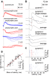
Figure 1. Polysynaptic Events in Postsynaptic Cells Initiated by Single Spikes of Human Pyramidal Cells
(A) Individual presynaptic (Pre) action potentials in human pyramidal cells evoke mono- and polysynaptic responses in postsynaptic (Post) pyramidal cells (red) and interneurons (blue). Ten traces showing monosynaptic EPSPs (top), monosynaptic EPSPs followed by polysynaptic IPSPs (middle), and polysynaptic IPSPs and EPSPs (bottom) with increasing onset latencies. Polysynaptic events occurred in 100% of trials in each of these four experiments. The timing of the presynaptic action potential is indicated by the dashed line. (B) Single action potentials in pyramid 1–initiated sequences of multiple events in simultaneously recorded pyramidal cells (pyramids 2 and 3). Disynaptic IPSPs occurred synchronously on the two postsynaptic cells. Disynaptic IPSPs were followed by presumably polysynaptic EPSPs and IPSPs in pyramid 2 and downstream IPSPs in pyramid 3. Blue and red dots indicate polysynaptic IPSP and EPSP onset times, respectively. (C) Scatter diagram of 54 consecutive sweeps in response to single presynaptic spikes. Blue and red dots correspond to the onset of individual IPSPs and EPSPs, respectively. (D) Double logarithmic correlation between the standard deviation (SD) and the mean of latency of polysynaptic postsynaptic potentials.
Figure 2
Figure 2. Relative Timing of Pairs of Polysynaptic Postsynaptic Potentials Detected in One or Several Postsynaptic Neurons within an Event Series Triggered by a Single Spike of Human Pyramidal Cells
(A) Temporally uncorrelated event sequences triggered by single presynaptic spikes: the latency of event 1 does not predict the timing of a different polysynaptic event (event 2). (B) Groups of postsynaptic potentials with temporally correlated event sequences: the latency of event 1 shifts together with that of event 2 relative to the trigger spike. Correlated latencies of pairs of polysynaptic potentials occur up to 35 ms after the single action potential initiating the series of network events. Colors correspond to different experiments. Insets, magnification of the boxed region near zero on the graph.
Figure 3
Figure 3. Sequential Polarity and Pharmacology of Single Pyramidal Spike–Triggered Polysynaptic Events in the Human Microcircuit
(A–C) Polysynaptic IPSPs are mediated by GABAA receptors and require the activation of AMPA-type glutamate receptors. (A) Monosynaptic connection between pyramidal cells 1 and 2. (B) A third pyramidal cell (pyramid 3) triggers an inhibitory response in pyramid 2, but the response has a longer latency than the monosynaptic connections shown in (A). The inhibitory response is sensitive to both gabazine and NBQX, showing the involvement of GABAA and AMPA receptors. (C) Light microscopic reconstruction of the three pyramidal cells (colors correspond to the panels [A and B]). Post, postsynaptic; Pre, presynaptic. (D and E) Polysynaptic EPSPs require preceding activation of GABAA receptor mediated event(s) in the network. (D) Single spikes in a presynaptic pyramidal cell (top) trigger polysynaptic IPSPs and EPSPs in a postsynaptic fast-spiking basket cell (middle). The polysynaptic IPSPs as well as polysynaptic EPSPs are sensitive to the GABAA receptor antagonist gabazine (bottom). (E) Temporal distribution of EPSPs (top) and IPSPs (bottom) recorded in experiments when single spikes triggered polysynaptic EPSPs. Monosynaptic EPSPs were followed exclusively by disynaptic IPSPs, and thus polysynaptic EPSPs had minimally trisynaptic latencies. Tri- and polysynaptic EPSPs and all IPSPs were gabazine sensitive as shown in panel (D).
Figure 4
Figure 4. Amplitude Distribution of Unitary EPSPs Arriving at Pyramidal Cells (Bottom) and Fast-Spiking Interneurons (Top) from Local Pyramidal Cells in Layers 2/3 of the Human Cortex
Note the unitary EPSPs of enormous amplitude selectively targeting fast-spiking interneurons. Inset, amplitude distribution of spontaneous EPSPs arriving at presynaptic human pyramidal cells (red) and postsynaptic interneurons (blue). The distribution of spontaneous EPSPs was shifted towards significantly higher amplitudes in interneurons relative to pyramidal cells.
Figure 5
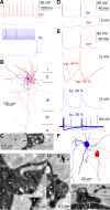
Figure 5. Spike-to-Spike Transmission from a Pyramidal Cell to a Basket Cell in the Human Neocortex
(A) Firing patterns of the pyramidal cell (pyr; red) and the basket cell (bc; blue) and their responses to hyperpolarizing current pulses. (B) Light microscopic reconstruction of the pyramidal cell (red) and the basket cell (blue). (C) Electron micrograph showing an axon terminal of the basket cell forming a synaptic junction (arrow) on the soma (s) of an unlabeled neuron. (D) Action potentials in the basket cell (top) elicit monosynaptic IPSPs in the pyramidal cell (bottom). (E) Action potentials in the pyramid (red traces) evoke EPSPs (44% of trials) and EPSPs eliciting action potentials (56% of trials) in the basket cell (blue traces). When postsynaptic spikes are triggered, IPSPs arriving back from the basket cell increase the amplitude of the afterhyperpolarization following the action potential of the pyramidal cell. (F) Route of the presynaptic pyramidal axon to the synapses formed on the dendrites of the basket cell. (F1–F3) Correlated light and electron microscopic identification of the three synaptic junctions (arrows on electron micrographs) between the axon (a) of the pyramidal cell and dendrites (d) of the basket cell. Numbering of synapses correspond to panel (F).
Figure 6
Figure 6. Human Axo-Axonic Cells Are Involved in Single Pyramidal Cell–Initiated Network Events
(A) Single action potentials in a presynaptic (Pre) pyramidal neuron (top) were effective in triggering postsynaptic (Post) action potentials with high-amplitude unitary responses in a postsynaptic axo-axonic cell (aac). The action potentials in the pyramidal neuron resulted in monosynaptic EPSPs in 65% of the trials eliciting second-order action potentials (middle), whereas in 35% of the trials, only subthreshold EPSPs were evoked (bottom). Note that postsynaptic, second-order action potentials in the axo-axonic cell were followed by occasional higher order spikes riding on presumably trisynaptic EPSPs. (B) Reconstruction of the somatodendritic (red) and axonal (black) arborization of the postsynaptic axo-axonic cell shown in panel (A). (C) Action potentials in a human axo-axonic cell trigger a sequence of polysynaptic events following a monosynaptic IPSP and a disynaptic EPSP in a postsynaptic pyramidal cell recorded with a low intracellular chloride concentration rendering all IPSPs hyperpolarizing in the two recorded cells. Note that downstream recurrent activity in the network triggers spikes in the axo-axonic cell. Responses of the pyramidal cell are shown as single sweeps (middle) and the average (bottom).
Figure 7
Figure 7. Polysynaptic Spike-to-Spike Coupling between Human Pyramidal Cells
Unitary action potentials in pyramidal cell 1 (top) elicited subthreshold, monosynaptic EPSPs in the postsynaptic (Post) basket cell (middle) and late-onset action potentials riding on polysynaptic EPSPs in pyramidal cell 2 (bottom). The peak of the presynaptic (Pre) action potential is projected by a dashed line for easier comparison of mono- and polysynaptic EPSP latencies.

Similar articles

Cited by

  • LRRC37B is a human modifier of voltage-gated sodium channels and axon excitability in cortical neurons.
    Libé-Philippot B, Lejeune A, Wierda K, Louros N, Erkol E, Vlaeminck I, Beckers S, Gaspariunaite V, Bilheu A, Konstantoulea K, Nyitrai H, De Vleeschouwer M, Vennekens KM, Vidal N, Bird TW, Soto DC, Jaspers T, Dewilde M, Dennis MY, Rousseau F, Comoletti D, Schymkowitz J, Theys T, de Wit J, Vanderhaeghen P. Libé-Philippot B, et al. Cell. 2023 Dec 21;186(26):5766-5783.e25. doi: 10.1016/j.cell.2023.11.028. Cell. 2023. PMID: 38134874 Free PMC article.
  • Remodeling of the axon initial segment after focal cortical and white matter stroke.
    Hinman JD, Rasband MN, Carmichael ST. Hinman JD, et al. Stroke. 2013 Jan;44(1):182-9. doi: 10.1161/STROKEAHA.112.668749. Epub 2012 Dec 11. Stroke. 2013. PMID: 23233385 Free PMC article.
  • Local connectivity and synaptic dynamics in mouse and human neocortex.
    Campagnola L, Seeman SC, Chartrand T, Kim L, Hoggarth A, Gamlin C, Ito S, Trinh J, Davoudian P, Radaelli C, Kim MH, Hage T, Braun T, Alfiler L, Andrade J, Bohn P, Dalley R, Henry A, Kebede S, Alice M, Sandman D, Williams G, Larsen R, Teeter C, Daigle TL, Berry K, Dotson N, Enstrom R, Gorham M, Hupp M, Dingman Lee S, Ngo K, Nicovich PR, Potekhina L, Ransford S, Gary A, Goldy J, McMillen D, Pham T, Tieu M, Siverts L, Walker M, Farrell C, Schroedter M, Slaughterbeck C, Cobb C, Ellenbogen R, Gwinn RP, Keene CD, Ko AL, Ojemann JG, Silbergeld DL, Carey D, Casper T, Crichton K, Clark M, Dee N, Ellingwood L, Gloe J, Kroll M, Sulc J, Tung H, Wadhwani K, Brouner K, Egdorf T, Maxwell M, McGraw M, Pom CA, Ruiz A, Bomben J, Feng D, Hejazinia N, Shi S, Szafer A, Wakeman W, Phillips J, Bernard A, Esposito L, D'Orazi FD, Sunkin S, Smith K, Tasic B, Arkhipov A, Sorensen S, Lein E, Koch C, Murphy G, Zeng H, Jarsky T. Campagnola L, et al. Science. 2022 Mar 11;375(6585):eabj5861. doi: 10.1126/science.abj5861. Epub 2022 Mar 11. Science. 2022. PMID: 35271334 Free PMC article.
  • HCN channels at the cell soma ensure the rapid electrical reactivity of fast-spiking interneurons in human neocortex.
    Szegedi V, Bakos E, Furdan S, Kovács BH, Varga D, Erdélyi M, Barzó P, Szücs A, Tamás G, Lamsa K. Szegedi V, et al. PLoS Biol. 2023 Feb 6;21(2):e3002001. doi: 10.1371/journal.pbio.3002001. eCollection 2023 Feb. PLoS Biol. 2023. PMID: 36745683 Free PMC article.
  • Developmental mechanisms underlying the evolution of human cortical circuits.
    Vanderhaeghen P, Polleux F. Vanderhaeghen P, et al. Nat Rev Neurosci. 2023 Apr;24(4):213-232. doi: 10.1038/s41583-023-00675-z. Epub 2023 Feb 15. Nat Rev Neurosci. 2023. PMID: 36792753 Free PMC article. Review.

References

    1. Thomson AM, Radpour S. Excitatory connections between CA1 pyramidal cells revealed by spike triggered averaging in slices of rat hippocampus are partially NMDA receptor mediated. Eur J Neurosci. 1991;3:587–601. - PubMed
    1. Miles R. Synaptic excitation of inhibitory cells by single CA3 hippocampal pyramidal cells of the guinea-pig in vitro. J Physiol. 1990;428:61–77. - PMC - PubMed
    1. Buhl EH, Halasy K, Somogyi P. Diverse sources of hippocampal unitary inhibitory postsynaptic potentials and the number of synaptic release sites. Nature. 1994;368:823–828. - PubMed
    1. Markram H, Lubke J, Frotscher M, Sakmann B. Regulation of synaptic efficacy by coincidence of postsynaptic APs and EPSPs. Science. 1997;275:213–215. - PubMed
    1. Povysheva NV, Gonzalez-Burgos G, Zaitsev AV, Kroner S, Barrionuevo G, et al. Properties of excitatory synaptic responses in fast-spiking interneurons and pyramidal cells from monkey and rat prefrontal cortex. Cereb Cortex. 2006;16:541–552. - PubMed

Publication types