. 2008 Nov;245(1-2):24-34.
doi: 10.1016/j.heares.2008.08.005. Epub 2008 Aug 19.

Over-expression of BDNF by adenovirus with concurrent electrical stimulation improves cochlear implant thresholds and survival of auditory neurons

Affiliations

Over-expression of BDNF by adenovirus with concurrent electrical stimulation improves cochlear implant thresholds and survival of auditory neurons

Jennifer A Chikar et al. Hear Res. 2008 Nov.

Abstract

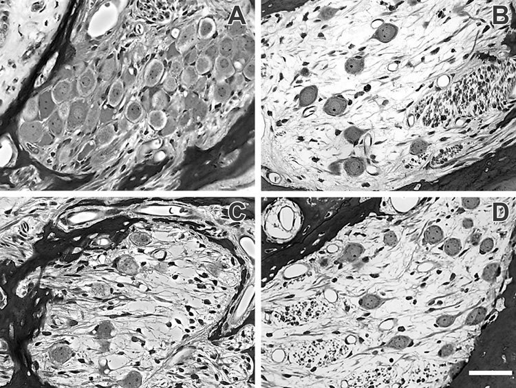
The survival of the auditory nerve in cases of sensorineural hearing loss is believed to be a major factor in effective cochlear implant function. The current study assesses two measures of cochlear implant thresholds following a post-deafening treatment intended to halt auditory nerve degeneration. We used an adenoviral construct containing a gene insert for brain-derived neurotrophic factor (BDNF), a construct that has previously been shown to promote neuronal survival in a number of biological systems. We implanted ototoxically deafened guinea pigs with a multichannel cochlear implant and delivered a single inoculation of an adenovirus suspension coding for BDNF (Ad.BDNF) into the scala tympani at the time of implantation. Thresholds to electrical stimulation were assessed both psychophysically and electrophysiologically over a period of 80 days. Spiral ganglion cell survival was analyzed at the 80 days time point. Compared to the control group, the Ad.BDNF treated group had lower psychophysical and electrophysiological thresholds as well as higher survival of spiral ganglion cells. Electrophysiological, but not psychophysical, thresholds correlated well with the density of spiral ganglion cells. These results indicate that the changes in the anatomy of the auditory nerve induced by the combination of Ad.BDNF inoculation and the electrical stimulation used for testing improved functional measures of CI performance.


Figures

Figure 1
Figure 1. Diagram of multichannel implant and electrode configurations
The banded electrodes are labeled A through F, where A is at the most apical (first inserted) end of the implant. MP = monopolar stimulation where the return electrode was a ground wire placed in the post-auricular muscle. BP = bipolar stimulation where the return electrode was immediately adjacent to the stimulating electrode. TP = tripolar stimulation where the two electrodes surrounding the stimulating electrode were return electrodes. All stimulation configurations are placed on arbitrary electrodes for illustration purposes.
Figure 2
Figure 2. Psychophysical detection thresholds averaged over time
Each column represents a different electrode configuration (A–B–C, A–B, B–G, C–D–E, D–E, D–G). Closed symbols represent averages over time of individual animal data points for each configuration. Open symbols represent the group average (n = 4) and error bars are +/− standard error of the mean (SEM). Circles represent animals that received Ad.BDNF inoculation and triangles indicate animals that received Ad.Empty inoculation. Differences between groups were assessed via Student’s t-test; (*) indicates p ≤ 0.05. P-values for each configuration are as follows: A–B–C: p = 0.06, A–B: p = 0.03, B–G: p = 0.61, C–D–E: p = 0.56, D–E: p = 0.38, D–G: p = 0.68.
Figure 3
Figure 3. Psychophysical detection thresholds over time, A–B configuration
Each data point represents the average of psychophysical detection thresholds collected in the preceding two-week period for the four animals in each experimental group. Most animals did not have valid thresholds in the first week following implantation; data therefore begin with the third week averages. Circles indicate Ad.BDNF treated animal averages and triangles indicate Ad.Empty treated animal averages at each time point. Error bars indicate +/− one standard deviation. A two-way repeated measures ANOVA revealed an effect of group (p = 0.02) but not a significant effect of time (p = 0.63) or interaction between time and group (p = 0.76). Post-hoc multiple pairwise comparisons gave the following p-values: 3 weeks = 0.21, 5 weeks = 0.07, 7 weeks = 0.09, 9 weeks = 0.06, 11 weeks = 0.06.
Figure 4
Figure 4. EABR thresholds averaged over time
Symbols and statistical tests as in Figure 2. P-values for each configuration: A–B: p = 0.002, B–G: p = 0.08, D–E: p = 0.03, D–G: p = 0.16.
Figure 5
Figure 5. EABR detection thresholds over time, A–B configuration
Each data point represents a group average of thresholds obtained at each time point indicated. Symbols as in Figure 3. A two-way repeated measures ANOVA revealed a significant interaction between group and time (p = 0.01), and (*) indicates a significant difference between groups was found at that time point. Post-hoc multiple pairwise comparisons gave the following p-values: 1 week = 0.47, 3 weeks = 0.03, 5 weeks = 0.02, 7 weeks = 0.03, 9 weeks = 0.03, 11 weeks = 0.04.
Figure 6
Figure 6. Light microscopy images of the organ of Corti structure of the basal turn
(A) represents the Ad.BDNF group, (B) represents the Ad.Empty group, and (C) represents the non-deafened, non-inoculated control group. There are no hair cells or supporting cells visible in (A) or (B), while inner hair cells (white arrow) and outer hair cells (black arrows) are observed in non-neomycin deafened ears (C). Scale bar = 50 µm, Adobe Photoshop ™ software was used to adjust contrast levels in all images.
Figure 7
Figure 7. Light microscopy images of the upper basal and middle turn of an Ad.BDNF (A, C) and an Ad.Empty (B, D) inoculated animal
The basal turn Rosenthal’s canal (A, B) showed a greater number of surviving SGCs in the Ad.BDNF treated ear (A) than the Ad.Empty cochlea (B). The middle turn Rosenthal’s canal (C, D) showed little difference between treatment groups in the number of surviving SGCs. Scale bar = 50 µm, Adobe Photoshop ™ software was used to adjust contrast levels in all images.
Figure 8
Figure 8. SGC density per cochlear turn for the Ad.BDNF and Ad.Empty groups
Symbols as in Figure 2. (*) indicates p ≤ 0.05, Student’s t-test p-values were: Lower Base: p = 0.10, Upper Base: p = 0.02, Middle: p = 0.42, Apex: p = 0.60.
Figure 9
Figure 9. Relationship between implant functional thresholds and number of surviving SGCs in (A) psychophysical testing and (B) electrophysiological testing
Psychophysical detection thresholds and EABR thresholds from the A–B configuration and SGC density from the upper basal turn are compared. Individual animal data points are plotted; circles represent animals that received Ad.BDNF inoculation and triangles indicate animals that received Ad.Empty inoculation. Lines represent linear regressions through all data points (r2 = 0.04 in A and r2 = 0.72 in B).

References

    1. Adamson CL, Reid MA, Davis RL. Opposite actions of brain-derived neurotrophic factor and neurotrophin-3 on firing features and ion channel composition of murine spiral ganglion neurons. J Neurosci. 2002;22:1385–1396. - PMC - PubMed
    1. Altschuler RA, Cho Y, Ylikoski J, Pirvola U, Magal E, Miller JM. Rescue and regrowth of sensory nerves following deafferentation by neurotrophic factors. Ann N Y Acad Sci. 1999;884:305–311. - PubMed
    1. Bowers WJ, Chen X, Guo H, Frisina DR, Federoff HJ, Frisina RD. Neurotrophin-3 transduction attenuates cisplatin spiral ganglion neuron ototoxicity in the cochlea. Mol Ther. 2002;6:12–18. - PubMed
    1. Colombo J, Parkins CW. A model of electrical excitation of the mammalian auditory-nerve neuron. Hear Res. 1987;31:287–311. - PubMed
    1. Di Polo A, Aigner LJ, Dunn RJ, Bray GM, Aguayo AJ. Prolonged delivery of brain-derived neurotrophic factor by adenovirus-infected muller cells temporarily rescues injured retinal ganglion cells. Proc Natl Acad Sci U S A. 1998;95:3978–3983. - PMC - PubMed

Publication types

MeSH terms

Substances