. 2008 Nov 7;283(45):31021-9.
doi: 10.1074/jbc.M805023200. Epub 2008 Sep 3.

Regulation of cyst wall protein promoters by Myb2 in Giardia lamblia

Affiliations

Regulation of cyst wall protein promoters by Myb2 in Giardia lamblia

Yu-Chang Huang et al. J Biol Chem. .

Abstract

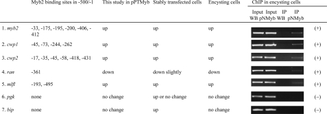
Myb family transcription factors are important in regulating cell proliferation, differentiation, and cell cycle progression. Giardia lamblia differentiates into infectious cysts to survive outside of the host. During encystation, genes encoding cyst wall proteins (CWPs) are coordinately induced. We have identified an encystation-induced Myb2 protein, which binds to the promoter regions of the cwp genes and myb2 itself in vitro. To elucidate the role of Myb2 in G. lamblia, we tested the hypothesis that Myb2 can activate encystation-induced genes. We found that overexpression of Myb2 resulted in an increase of expression of CWP1 at both protein and mRNA levels. Interestingly, the Myb2-overexpressing trophozoites had increased capability to differentiate into cysts. In cotransfection assays, Myb2 was able to transactivate the cwp promoters and its own promoter in vivo, suggesting that its gene can be positively autoregulated. Moreover, deletion of the N- or C-terminal domain resulted in a decrease of transactivation and autoregulation function of Myb2. We also found that the promoter of a newly identified encystation-induced gene, the giardial myeloid leukemia factor-like gene, has the Myb2 binding sites and that its mRNA levels were increased by Myb2 overexpression. Chromatin immunoprecipitation assays confirmed that Myb2 was bound to the promoters with its binding sites. Transfection of the myb2 antisense construct reduced the levels of the cwp1 transcripts and cyst formation. Our results suggest that Myb2 is a potent transactivator of the cwp genes and other endogenous genes and plays an important role in G. lamblia differentiation into cysts.


Figures

FIGURE 1.
FIGURE 1.
Induction of cwp1 and cwp2 gene expression in the Myb2-overexpressing cell line. A, diagrams of the 5′-Δ5N-Pac and pPTMyb2 plasmid. The pac gene (open box) is under the control of the 5′- and 3′-flanking regions of the gdh gene (striated box). In construct pPTMyb2, the myb2 gene is under the control of the constitutively expressed α2-tubulin promoter (striped box) and 3′-flanking region of the ran gene (dotted box). The filled box indicates the coding sequence of the AU1 epitope tag. B, Northern blot analysis of gene expression in the Myb2-overexpressing cell line. Total RNA blots made from 5′-Δ5N-Pac or pPTMyb2 cell cultures during vegetative growth were hybridized with specific gene probes as indicated (upper panels). Ribosomal RNA loading controls are in the bottom panel. Representative results are shown. The numbers show the relative activity, which reflects expression relative to that in controls.
FIGURE 2.
FIGURE 2.
Localization of CWP1 in the Myb2-overexpressing cell line. A, overexpression of Myb2 increased the levels of cyst formation. 5′-Δ5N-Pac and pPTMyb2 stable transfectants were cultured in growth medium to late log/early stationary phase. Cyst count was performed on these late log/early stationary phase cultures (1.5 × 10⁶ cells/ml). The sum of total cysts is expressed as relative expression level over control. Values are shown as means ± S.E. B, overexpression of Myb2 increased the numbers of CWP1 positive staining cells. The 5′Δ5N-Pac and pPTMyb2 stable transfectants were cultured in growth medium to late log/early stationary phase and then subjected to immunofluorescence assays. The endogenous CWP1 protein was detected by anti-CWP1 antibody. The number of CWP1-positive staining cells were counted and expressed as relative expression level over control. Values are shown as means ± S.E. C and D, localization of CWP1 and Myb2 in the same cell. The endogenous CWP1 protein and vector-expressed Myb2-AU1 protein were detected by anti-CWP1 and anti-AU1 antibodies. The left panel shows that the CWP1 protein is localized to the ESVs. The middle panel shows that the Myb2-AU1 protein is localized to the nuclei and slightly in the cytoplasm. The right panel shows the merged image. The CWP1 protein was stained in the ESVs of all of Myb2-AU1-positive stained cells (data not shown), and two cells are shown in C and D.
FIGURE 3.
FIGURE 3.
Transactivation by Myb2 through its own target sequence. In construct pNTMyb2, the myb2 gene is under the control of the constitutively expressed α2-tubulin promoter (striped box) and 3′-flanking region of the ran gene (dotted box). The filled box indicates the coding sequence of the AU1 epitope tag. In the pPW1, pPC35, or pPM5 reporter construct, the luciferase gene (luc+, open box) is under the control of the cwp1, cwp3, or myb2 promoter as shown. In the -42/+3 reporter construct, the luciferase gene (luc, open box) is under the control of the g6pi-b promoter deletion lacking the Myb2 binding site. In the 4s/-42/+3 reporter construct, four Myb2 binding sequences (open boxes) were inserted upstream of the g6pi-b promoter deletion lacking the Myb2 binding site. The neo or pac gene (open box) is under the control of the 5′- and 3′-flanking regions of the ran (dotted box) or gdh gene (striated box). Specific cell lines were produced by the stable co-transfection of reporter construct and the effector construct pNTMyb2. The corresponding control cell lines were produced by the stable co-transfection of reporter construct and the pRANneo construct. Luciferase activity was measured in vegetative cells as described under “Experimental Procedures.” The activity of co-transfectants (+pNTMyb2) relative to the corresponding control cell line (+pRANneo) is presented. Values are shown as means ± S.E. in the right panel.
FIGURE 4.
FIGURE 4.
Localization of Myb2 and its deletion derivatives. A, diagrams of the plasmids for Myb2 deletion mapping. Each plasmid contains a neo gene (open box) under the control of the 5′- and 3′-flanking regions of the ran gene (dotted box). Plasmid pNMyb contains the myb2 gene (open boxes) under the control of the myb2 its own promoter (striped box) and 3′-flanking region of the ran gene (dotted box). The filled box indicates the coding sequence of the AU1 epitope tag. The Myb repeats are indicated as open boxes labeled Myb. The specific transfectants were cultured in growth medium (Veg) or encystation medium for 24 h (Enc) and then subjected to immunofluorescence analysis, using anti-AU1 antibody for detection. The localization of Myb2 in nuclei (N) or cytosol (C) is summarized in the right panel. “-” denotes negative staining. B, alignment of the amino acid sequences of the giardial Myb2 and P. putida dehRI or S. cerevisiae Rlr1p. Specific sequence similarity search was performed against the GenBank™ data base on the NCBI Web site using the BLASTP algorithm (50). This search identified similarity of giardial Myb2 to P. putida dehRI or S. cerevisiae Rlr1p in the GenBank™ data base. Numbers indicate positions of the residues relative to the first amino acid. Plus (+) and hyphens indicate conserved amino acids and gaps in the respective proteins, respectively.
FIGURE 5.
FIGURE 5.
Mapping of the Myb2 transactivation domain. A, analysis of Myb2 deletion mutants. The specific transfectants were cultured in encystation medium for 24 h (Enc) and then subjected to Western blot analysis, using anti-AU1 antibody for detection. Coomassie Blue-stained total protein loading control is shown below. B, analysis of cwp1 gene expression. Total RNA was harvested from specific transfectants cultured in encystation medium for 24 h. Northern blots were hybridized with the cwp1 and myb2 gene probes (upper panels). Ribosomal RNA loading controls are in the bottom panels. Representative results are shown. The numbers show the relative activity, which reflects expression relative to that in controls. Full-length myb2 transcripts (shown by arrows) and deletion mutant transcripts (shown by arrowheads) were detected in the pNMybΔC and pNMybΔN transfectants. Only the relative activity of the full-length myb2 transcripts (shown by arrows) was shown for the pNMybΔC and pNMybΔN transfectants.
FIGURE 6.
FIGURE 6.
Mutation analysis of the Myb2 binding site in the myb2 promoter region. In the pNM5 construct, the luciferase gene (luc+, open box) is under the control of the myb2 promoter and 3′-flanking region of the ran gene (dotted box). The neo gene is under the control of the 5′- and 3′-flanking regions of the ran gene (dotted box). In the pNM5m construct, the mutated Myb2 binding site is indicated by a filled box. After stable transfection with these constructs, luciferase activity was measured in vegetative cells and 24 h encysting cells as described under “Experimental Procedures.” Values are shown as means ± S.E. in the right panel. The induction ratio was obtained by dividing the activity in the encysting cells by the activity in the vegetative cells of each construct.
FIGURE 7.
FIGURE 7.
Summary of the results from search of Myb2 binding sites and ChIP assays. The 500 bp of the 5′-flanking regions of endogenous genes were searched for Myb2 binding sites CTACAG, CAACAG, CTACTG, CTACAC on the coding strand or noncoding strand. The 3′-ends of the Myb2 binding sites are numbered relative to the translation initiation site (+1). The pNMyb cell line expressing the AU1-tagged Myb2 (Fig. 4) and non-transfected WB cells as the control cell line were cultured in encystation medium for 24 h and then subjected to ChIP assays. Anti-AU1 antibody conjugated to beads was used to assess binding of Myb2-AU1 to endogenous gene promoters. Immunoprecipitated chromatin was analyzed by PCR using primers that amplify the 5′-flanking region of specific genes. Approximately the same amount of PCR product was obtained from input DNA in the pNMyb cell line and the control cell line. At least three independent experiments were performed. Representative results are shown. Immunoprecipitated products of Myb2-AU1 yielded more PCR products of myb2, cwp1, cwp2, ran, and mlfl promoters in the pNMyb cell line relative to the control cell line, indicating that Myb2-AU1 was bound to these promoters (+).
FIGURE 8.
FIGURE 8.
Transfection of the myb2 antisense construct reduced the levels of the cwp1 transcripts and cyst formation. A, diagrams of the myb2 antisense construct. A 1590-bp PCR fragment of the myb2 gene was cloned in the opposite (antisense) orientation to its own promoter into the pNM5 (Fig. 6). The resulting construct, pNMyb2as, contains full-length coding sequences complementary to the mRNA sequences of the myb2 gene. B, transfection of the myb2 antisense construct reduced the levels of cyst formation. Wild-type non-transfected WB C6 cells, pNM5 and pNMyb2as stable transfectants were cultured in encystation medium for 48 h and then subjected to cyst count. The sum of total cysts is expressed as relative expression level over control. Values are shown as means ± S.E. C, transfection of the myb2 antisense construct reduced the cwp1 transcript levels. Total RNA blots made from vegetative non-transfected WB C6 cells, pNM5 or pNMyb2as stable transfectants were hybridized with specific probes as indicated (upper panels). Ribosomal RNA loading controls are in the bottom panel. Representative results are shown. The numbers show the relative activity, which reflects expression relative to that in controls.

Similar articles

Cited by

References

    1. Marshall, M. M., Naumovitz, D., Ortega, Y., and Sterling, C. R. (1997) Clin. Microbiol. Rev. 10 67-85 - PMC - PubMed
    1. Gillin, F. D., and Reiner, D. S. (1996) Annu. Rev. Microbiol. 50 679-705 - PubMed
    1. Eichinger, D. (2001) Curr. Opin. Microbiol. 4 421-426 - PubMed
    1. Wolfe, M. S. (1992) Clin. Microbiol. Rev. 5 93-100 - PMC - PubMed
    1. Adam, R. D. (2001) Clin. Microbiol. Rev. 14 447-475 - PMC - PubMed

Publication types

MeSH terms

Substances

Associated data

LinkOut - more resources