. 2008 Nov 7;283(45):30766-71.
doi: 10.1074/jbc.M805353200. Epub 2008 Sep 2.

Entrapment of carbon dioxide in the active site of carbonic anhydrase II

Affiliations

Entrapment of carbon dioxide in the active site of carbonic anhydrase II

John F Domsic et al. J Biol Chem. .

Abstract

The visualization at near atomic resolution of transient substrates in the active site of enzymes is fundamental to fully understanding their mechanism of action. Here we show the application of using CO(2)-pressurized, cryo-cooled crystals to capture the first step of CO(2) hydration catalyzed by the zinc-metalloenzyme human carbonic anhydrase II, the binding of substrate CO(2), for both the holo and the apo (without zinc) enzyme to 1.1A resolution. Until now, the feasibility of such a study was thought to be technically too challenging because of the low solubility of CO(2) and the fast turnover to bicarbonate by the enzyme (Liang, J. Y., and Lipscomb, W. N. (1990) Proc. Natl. Acad. Sci. U. S. A. 87, 3675-3679). These structures provide insight into the long hypothesized binding of CO(2) in a hydrophobic pocket at the active site and demonstrate that the zinc does not play a critical role in the binding or orientation of CO(2). This method may also have a much broader implication for the study of other enzymes for which CO(2) is a substrate or product and for the capturing of transient substrates and revealing hydrophobic pockets in proteins.


Figures
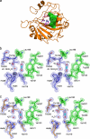
FIGURE 1.
FIGURE 1.
hCAII structure. a, overall view, showing the hydrophilic (magenta stick representation) and hydrophobic (green surface representation) sides of the active site. The active site zinc is shown in purple with the waters of the proton wire shown as small, red spheres. b and c, a close-up stereo view of the active site showing the position of bound CO2 in holo- (b) and apohCAII (c). Electron density of the active site amino acids and WI (σ-weighted 2Fo - Fc Fourier map contoured at 2.25 σ) and CO2 (σ-weighted Fo - Fc Fourier map contoured at 2.25 σ). The figure was created using PyMOL.
FIGURE 2.
FIGURE 2.
Second CO2 binding site. a, surface representation showing the separation of the active site (green) and non-catalytic (pink) CO2 binding pockets. b, close-up view of the CO2 binding. Note the conformational change in Phe-226 (red = unbound, green = CO2-bound holohCAII). The electron density is a σ-weighted 2Fo - Fc Fourier map contoured at 1.5 σ. The figure was created using PyMOL.
FIGURE 3.
FIGURE 3.
Glycerol binding sites in CO2-bound hCAII structures. a, ordered glycerol molecule located at mouth of the active site in the holo enzyme. b and c, second ordered glycerol observed bound on the surface of the holo (b) and apo (c) structures. The electron density maps are 2Fo - Fc Fourier map contoured at 1.0 σ. The figure was made using PyMOL.
FIGURE 4.
FIGURE 4.
Active site. a, superposition of unbound holo- (13), CO2-bound holo-, and bicarbonate-bound T200H hCAII (32). The binding modes of both the CO2 substrate and the formula image product are similar, with the substrate favoring the hydrophobic side (green) and product favoring the hydrophilic side (orange). Note that the deep water (WDW, gray sphere) is displaced and a new water occupies the area between the side chain of Thr-200 and CO2 (WI, cyan) upon CO2 binding. The CO2 is orientated so that the carbon is primed for the nucleophilic attack by the zinc-bound hydroxide (orange sphere). b, a superposition of the V143Y variant of hCAII (9, 27). Note that the side chain of Tyr-143 (white) acts as a steric block to the CO2 binding site. The figure was created using PyMOL.
FIGURE 5.
FIGURE 5.
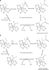
Proposed mechanisms of hCAII catalysis. a and b, Lipscomb (32) mechanism (a) and Lindskog (33) (b) mechanism.
FIGURE 6.
FIGURE 6.
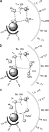
Proposed catalytic mechanism of hCAII. A schematic representation of three discrete stages of the catalytic cycle is shown. a, unbound. Note the presence of deep water (WDW). b, CO2 bound. Note the displacement of WDW and the hydrogen bond between the substrate and backbone amide of Thr-199. c, the formation of bicarbonate. The figure was created using ChemDraw 11.0 (available from CambridgeSoft).

References

    1. Liang, J. Y., and Lipscomb, W. N. (1990) Proc. Natl. Acad. Sci. U. S. A. 87 3675-3679 - PMC - PubMed
    1. Davenport, H. W. (1984) Ann. N Y Acad. Sci. 429 4-9 - PubMed
    1. Chegwidden, W. R., Carter, N. D., and Edwards, Y. H. (2000). The Carbonic Anhydrases, New Horizons, Birkhauser Verlag Basel, Switzerland
    1. Christianson, D. W., and Fierke, C. A. (1996) Acc. Chem. Res. 29 331-339
    1. Xu, Y., Feng, L., Jeffrey, P. D., Shi, Y., and Morel, F. M. (2008) Nature 452 56-61 - PubMed

Publication types

MeSH terms

LinkOut - more resources