A pilot study of compositional analysis of the breast and estimation of breast mammographic density using three-dimensional T1-weighted magnetic resonance imaging
- PMID: 18768492
- PMCID: PMC2582975
- DOI: 10.1158/1055-9965.EPI-07-2547
A pilot study of compositional analysis of the breast and estimation of breast mammographic density using three-dimensional T1-weighted magnetic resonance imaging
Abstract
Purpose: A method and computer tool to estimate percentage magnetic resonance (MR) imaging (MRI) breast density using three-dimensional T(1)-weighted MRI is introduced, and compared with mammographic percentage density [X-ray mammography (XRM)].
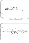
Materials and methods: Ethical approval and informed consent were obtained. A method to assess MRI breast density as percentage volume occupied by water-containing tissue on three-dimensional T(1)-weighted MR images is described and applied in a pilot study to 138 subjects who were imaged by both MRI and XRM during the Magnetic Resonance Imaging in Breast Screening study. For comparison, percentage mammographic density was measured from matching XRMs as a ratio of dense to total projection areas scored visually using a 21-point score and measured by applying a two-dimensional interactive program (CUMULUS). The MRI and XRM percent methods were compared, including assessment of left-right and interreader consistency.
Results: Percent MRI density correlated strongly (r = 0.78; P < 0.0001) with percent mammographic density estimated using Cumulus. Comparison with visual assessment also showed a strong correlation. The mammographic methods overestimate density compared with MRI volumetric assessment by a factor approaching 2.
Discussion: MRI provides direct three-dimensional measurement of the proportion of water-based tissue in the breast. It correlates well with visual and computerized percent mammographic density measurements. This method may have direct application in women having breast cancer screening by breast MRI and may aid in determination of risk.
Figures


References
References in Appendices
-
- Brown J, Buckley D, Coulthard A. Magnetic resonance imaging screening in women at genetic risk of breast cancer: imaging and analysis protocol for the UK multi-centre study. Magnetic Resonance Imaging. 2000;18:765–776. - PubMed
-
- Soltanian-Zadeh H, Windham JP. Optimal Processing of Brain MRI. In: Stricland RN, editor. Image-Processing Techniques for Tumor Detection. Marcel Dekker, Inc; 2002.
-
- Khazen M, Warren R, Leach MO. Automated algorithm and a computer tool for identifying tissues contributing to mammographic density using 3D T1-weighted dynamic contrast enhanced MRI of the breast (abstr); Radiological Society of North America scientific assembly and annual meeting program; Oak Brook, IL: Radiological Society of North America; 2005. p. 866.
-
- Khazen M, Warren R, Leach MO. Magnetic Resonance Imaging of the Breast, Analysis and Review (MRIBview): An Image Processing Tool for Structural, Compositional, and Functional Analysis of the Breast, and Computer-assisted Detection (CAD) of Breast Cancer (abstr); Radiological Society of North America scientific assembly and annual meeting program; Oak Brook, IL: Radiological Society of North America; 2006. p. 785.
-
- Khazen M, D'Arcy J, Collins D, Padhany A, Walker-Samuel S, Leach MO. Magnetic Resonance Beast Imaging Analysis and Review (MRIBview) – Specialized Image Processing Workstation for DCE-MRI of the Breast (abstr); Radiological Society of North America scientific assembly and annual meeting program; Oak Brook, IL: Radiological Society of North America; 2004. p. 821.
References
-
- McCormack VA, Dos SS,I. Breast density and parenchymal patterns as markers of breast cancer risk: a meta-analysis. Cancer Epidemiol Biomarkers Prev. 2006;15:1159–69. - PubMed
-
- Byng JW, Yaffe MJ, Jong RA, et al. Analysis of mammographic density and breast cancer risk from digitized mammograms. Radiographics. 1998;18:1587–98. - PubMed
-
- Poon CS, Bronskill MJ, Henkelman RM, Boyd NF. Quantitative magnetic resonance imaging parameters and their relationship to mammographic pattern. J Natl Cancer Inst. 1992;84:777–81. - PubMed
-
- Lee NA, Rusinek H, Weinreb J, et al. Fatty and fibroglandular tissue volumes in the breasts of women 20-83 years old: comparison of X-ray mammography and computer-assisted MR imaging. AJR Am J Roentgenol. 1997;168:501–6. - PubMed
Publication types
MeSH terms
Grants and funding
LinkOut - more resources
Full Text Sources
Other Literature Sources
Medical

