. 2008 Oct;14(10):2013-29.
doi: 10.1261/rna.1027208. Epub 2008 Sep 3.

Toward predicting self-splicing and protein-facilitated splicing of group I introns

Affiliations

Toward predicting self-splicing and protein-facilitated splicing of group I introns

Quentin Vicens et al. RNA. 2008 Oct.

Abstract

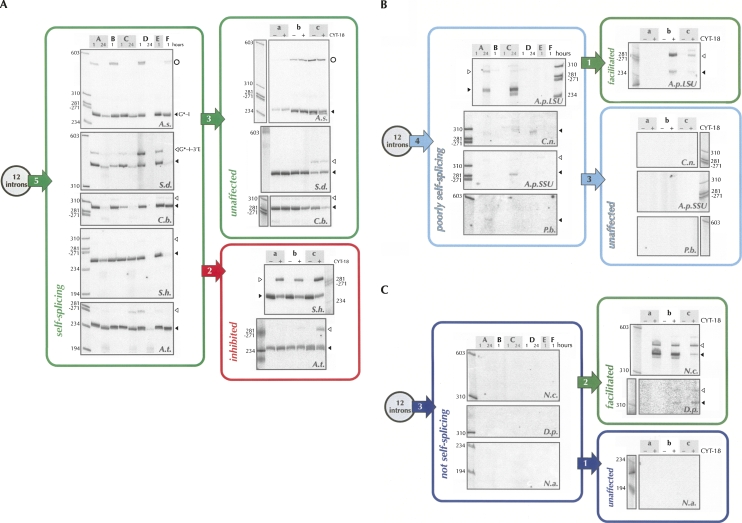
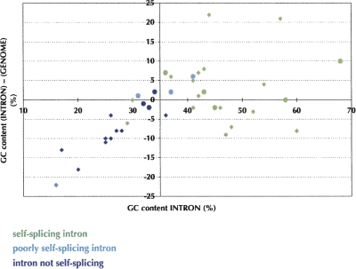
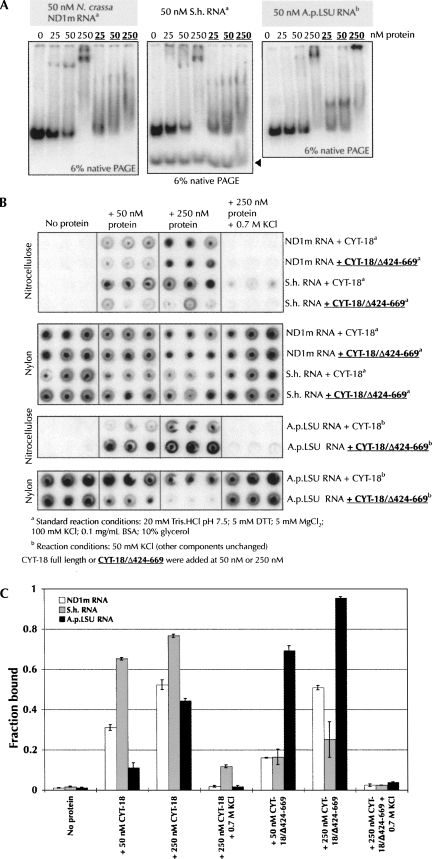
In the current era of massive discoveries of noncoding RNAs within genomes, being able to infer a function from a nucleotide sequence is of paramount interest. Although studies of individual group I introns have identified self-splicing and nonself-splicing examples, there is no overall understanding of the prevalence of self-splicing or the factors that determine it among the >2300 group I introns sequenced to date. Here, the self-splicing activities of 12 group I introns from various organisms were assayed under six reaction conditions that had been shown previously to promote RNA catalysis for different RNAs. Besides revealing that assessing self-splicing under only one condition can be misleading, this survey emphasizes that in vitro self-splicing efficiency is correlated with the GC content of the intron (>35% GC was generally conductive to self-splicing), and with the ability of the introns to form particular tertiary interactions. Addition of the Neurospora crassa CYT-18 protein activated splicing of two nonself-splicing introns, but inhibited the second step of self-splicing for two others. Together, correlations between sequence, predicted structure and splicing begin to establish rules that should facilitate our ability to predict the self-splicing activity of any group I intron from its sequence.


Figures

FIGURE 1.
FIGURE 1.
Mechanism of group I intron splicing. The intron-containing products that are labeled upon addition of [α-32P]GTP to the reaction mix, and hence visible on polyacrylamide gels, are shown in black, while the unlabeled precursor RNA and the released exons are shown in white.
FIGURE 2.
FIGURE 2.
Self-splicing and protein-facilitated splicing of the 12 group I introns studied. (A) Results of the activity assays of the self-splicing introns (green). Introns that are inhibited upon CYT-18 binding are circled in red. (B) Results of the activity assays of the poorly self-splicing introns (cyan). (C) Results of the activity assays of the introns that are not self-splicing (dark blue). Intron abbreviations are defined in Table 1. Times of incubation are 1 h and 24 h. The numbers in the arrows indicate the total number of introns that followed each scenario. The previously uncharacterized circular product from the self-splicing reaction of the A.s. intron (indicated by an open circle) will be described elsewhere.
FIGURE 3.
FIGURE 3.
Native gel electrophoresis for six introns. Pre, precursor; G-I, excised intron with GMP at its 5′ end; LE, ligated exons. The asterisk denotes the following modification of condition E: 0.4 M NH4Oac, 10 mM Mg(OAc)2. GMP was used in place of GTP in this particular series of experiments.
FIGURE 4.
FIGURE 4.
Correlation between self-splicing activity and the GC contents of 38 introns and of their host genomes. Introns from both this work (circles) and the literature (diamonds) are shown (Supplemental material). Same color code as in Figure 2.
FIGURE 5.
FIGURE 5.
Interaction network diagrams for three representative group I introns. Base pairing within long range interactions and structural motifs is shown using the nomenclature of Leontis and Westhof (2001). Circles denote interactions involving the Watson-Crick face, squares the Hoogsteen face, and triangles the sugar edge. A question mark on the A.p.LSU diagram indicates the ambiguous topology of the J5/5a junction (residues 100% conserved in the P5ab extensions of the A.p.LSU and A.p.SSU introns are shown in red). The self-splicing behavior is indicated in the same color code as in Figure 2.
FIGURE 6.
FIGURE 6.
Binding assays of the full-length and C-terminally truncated CYT-18 proteins to the ND1m, S.h., and A.p.LSU introns. (A) Gel shift assays with 50 nM RNA and increasing concentrations (0, 25 nM, 50 nM, and 250 nM) of CYT-18 full length or truncated (bold underlined). In this and subsequent panels, CYT-18 binding and protein-facilitation assays were carried out in standard conditions for the ND1m and S.h. introns (20 mM Tris-HCl at pH 7.5, 5 mM DTT, 5 mM MgCl2, 100 mM KCl, 0.1 mg/mL BSA, 10% glycerol) and at 50 mM KCl for the A.p.LSU intron (other components unchanged). The black arrow indicates the linearized S.h. intron. (B) Filter binding assays upon addition of increasing concentrations (0, 50 nM, and 250 nM) of CYT-18 full length or tuncated (bold underlined). Experiments were performed in triplicate. (C) Quantitation of the filter binding experiments shown in panel B. The error bars indicate the standard deviation calculated from the triplicate experiments shown in B.
FIGURE 7.
FIGURE 7.
Facilitation or inhibition of splicing upon addition of the CYT-18 protein. (A) Time course self-splicing experiment (5, 20, and 60 min time points) of the S.h. intron in the presence of increasing concentrations of the CYT-18 protein (onefold and fivefold of the RNA concentration). (B) Comparison of the effects of the addition of full length CYT-18 or of C-terminally truncated CYT-18 on the self-splicing activity of the N.c., S.h., and A.p.LSU introns. (C) Product ratios obtained in the presence of full length or C-terminally truncated CYT-18 protein. The error bars indicate the standard deviation calculated from three independent experiments.

References

    1. Adams, P.L., Stahley, M.R., Kosek, A.B., Wang, J., Strobel, S.A. Crystal structure of a self-splicing group I intron with both exons. Nature. 2004;430:45–50. - PubMed
    1. Akins, R.A., Lambowitz, A.M. A protein required for splicing group I introns in Neurospora mitochondria is mitochondrial tyrosyl-tRNA synthetase or a derivative thereof. Cell. 1987;50:331–345. - PubMed
    1. Baer, M.F., Arnez, J.G., Guerrier-Takada, C., Vioque, A., Altman, S. Preparation and characterization of RNase P from Escherichia coli . Methods Enzymol. 1990;181:569–582. - PubMed
    1. Baird, N.J., Srividya, N., Krasilnikov, A.S., Mondragon, A., Sosnick, T.R., Pan, T. Structural basis for altering the stability of homologous RNAs from a mesophilic and a thermophilic bacterium. RNA. 2006;12:598–606. - PMC - PubMed
    1. Beckert, B., Nielsen, H., Einvik, C., Johansen, S.D., Westhof, E., Masquida, B. Molecular modelling of the GIR1 branching ribozyme gives new insight into evolution of structurally related ribozymes. EMBO J. 2008;27:667–678. - PMC - PubMed

Publication types