. 2008 Sep 3;28(36):8908-13.
doi: 10.1523/JNEUROSCI.1526-08.2008.

Midbrain dopamine neurons: projection target determines action potential duration and dopamine D(2) receptor inhibition

Affiliations

Midbrain dopamine neurons: projection target determines action potential duration and dopamine D(2) receptor inhibition

Elyssa B Margolis et al. J Neurosci. .

Abstract

Broad action potentials (APs) and dopamine (DA) D(2) receptor (D(2)R)-mediated inhibition are widely used to identify midbrain DA neurons. However, when these measures are taken alone they do not predict DA content in ventral tegmental area (VTA) neurons. In fact, some VTA neuronal properties correlate better with projection target than neurotransmitter content. Here we report that amygdala (AMYG)-projecting VTA DA neurons have brief APs and lack D(2)R agonist (quinpirole; 1 microM) autoinhibition. However, they are hyperpolarized by both the GABA(B) agonist baclofen (1 microM) and the kappa-opioid receptor agonist U69593 [(+)-(5alpha,7alpha,8beta)-N-methyl-N-[7-(1-pyrrolidinyl)-1-oxaspiro[4.5]dec-8-yl]benzeneacetamide; 1 microM]. Furthermore, we show that accurate prediction of DA content in VTA neurons is possible when the projection target is known: in both nucleus accumbens- and AMYG-projecting neural populations, AP durations are significantly longer in DA than non-DA neurons. Among prefrontal cortex-projecting neurons, quinpirole sensitivity, but not AP duration, is a predictor of DA content. Therefore, in the VTA, AP duration and inhibition by D(2)R agonists may be valid markers of DA content in neurons of known projection target.


Figures

Figure 1.
Figure 1.
Action potential duration is correlated with D2R agonist inhibition in VTA neurons. Spontaneously firing VTA neurons from uninjected animals were tested for responses to bath-applied 1 μm quinpirole. Open symbols represent neurons in which TH content was not determined. “a” indicates the means ± SEM among neurons significantly inhibited by quinpirole, and “b” indicates the means ± SEM among neurons not inhibited by quinpirole.
Figure 2.
Figure 2.
Amygdala-projecting VTA DA neurons have short duration action potentials. A, Example APs collected from neurons that project to the AMYG, PFC, or NAc with the corresponding immunocytochemistry. Neurons were labeled by the retrograde marker DiI (red), filled with biocytin during the recording (green), and immunocytochemically identified as TH(+) (blue). B, The distribution of AP durations in confirmed TH(+) neurons (top), Ih(+), TH(−) neurons (middle), and Ih(−) neurons (bottom) for each projection target. Rectangles represent the 25th and 75th percentiles of each sample, the solid black vertical lines indicate the means, and the broken gray line at 1.87 ms is the longest AP duration recorded in an NAc-projecting TH(−) neuron.
Figure 3.
Figure 3.
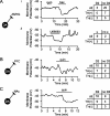
Amygdala-projecting VTA neurons are not hyperpolarized by D2R activation. Ai, Example AMYG-projecting TH(+) VTA neuron: 1 μm quinpirole (quin) application did not modulate the membrane potential; however, 1 μm of the GABAB receptor agonist baclofen (bac) hyperpolarized the cell. Aii, Example AMYG-projecting neuron hyperpolarized by the KOP-R agonist U69593 (1 μm). B, C, Example PFC-projecting (B) and NAc-projecting (C) TH(+) VTA neurons hyperpolarized by 1 μm quinpirole. Tables indicate the number of neurons hyperpolarized or not in each projection.
Figure 4.
Figure 4.
AP duration identifies TH(+) neurons in AMYG- and NAc-projecting VTA populations. A, A median split of the AP durations was made within each projection population among immunocytochemically identified neurons. Among AMYG- and NAc-projecting neurons, the probability is high that a neuron with an AP duration greater than the median is TH(+). AP duration does not correlate with TH content among PFC-projecting neurons. B, AP duration histograms of all recorded neurons for each projection. Lighter bars indicate durations less than the median. C, D2R inhibition is a better predictor of TH content among PFC-projecting neurons.

References

    1. Aghajanian GK, Bunney BS. Pharmacological characterization of dopamine “autoreceptors” by microiontophoretic single-cell recording studies. Adv Biochem Psychopharmacol. 1977;16:433–438. - PubMed
    1. Alderson HL, Latimer MP, Winn P. Intravenous self-administration of nicotine is altered by lesions of the posterior, but not anterior, pedunculopontine tegmental nucleus. Eur J Neurosci. 2006;23:2169–2175. - PubMed
    1. Bayer HM, Glimcher PW. Midbrain dopamine neurons encode a quantitative reward prediction error signal. Neuron. 2005;47:129–141. - PMC - PubMed
    1. Bayer VE, Pickel VM. Ultrastructural localization of tyrosine hydroxylase in the rat ventral tegmental area: relationship between immunolabeling density and neuronal associations. J Neurosci. 1990;10:2996–3013. - PMC - PubMed
    1. Bunney BS, Walters JR, Roth RH, Aghajanian GK. Dopaminergic neurons: effect of antipsychotic drugs and amphetamine on single cell activity. J Pharmacol Exp Ther. 1973a;185:560–571. - PubMed

Publication types

MeSH terms