. 2008 Sep 3;28(36):9055-65.
doi: 10.1523/JNEUROSCI.1424-08.2008.

Sex-specific programming of offspring emotionality after stress early in pregnancy

Affiliations

Sex-specific programming of offspring emotionality after stress early in pregnancy

Bridget R Mueller et al. J Neurosci. .

Abstract

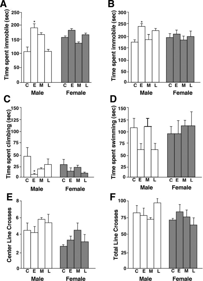
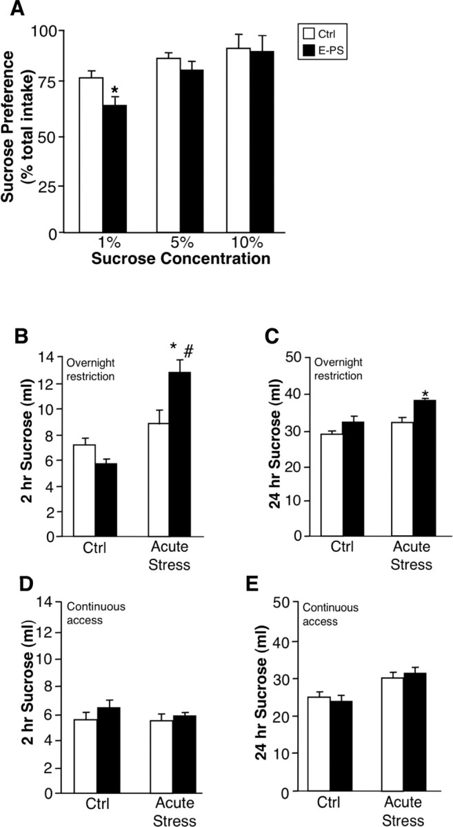
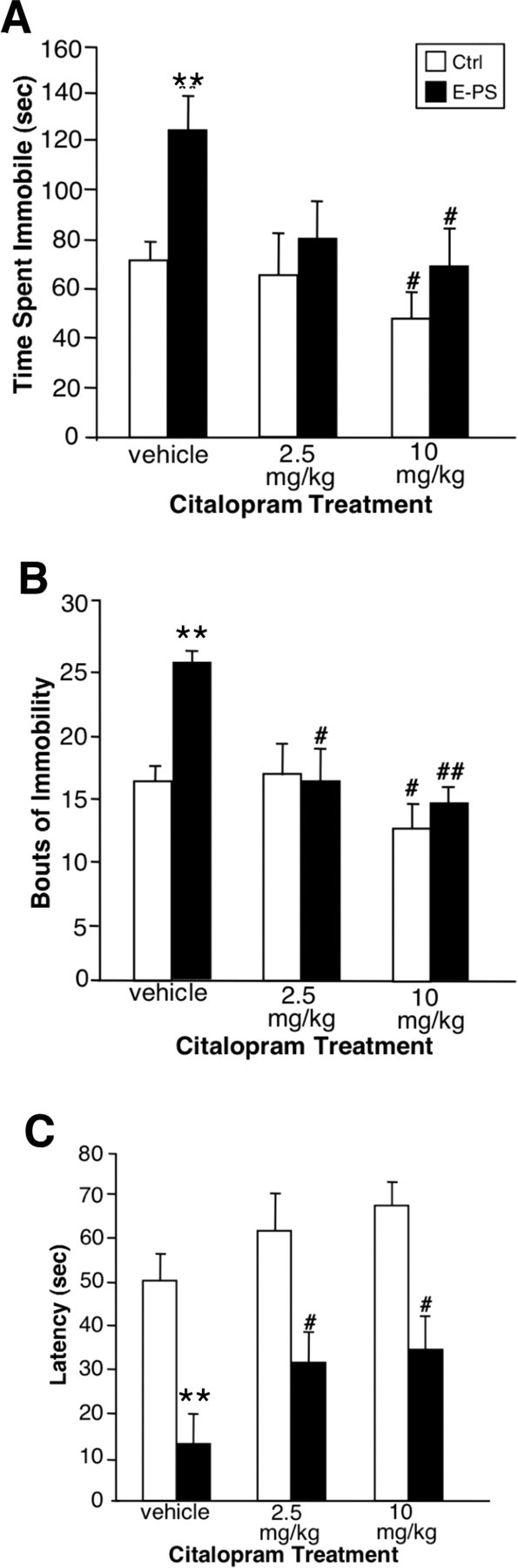
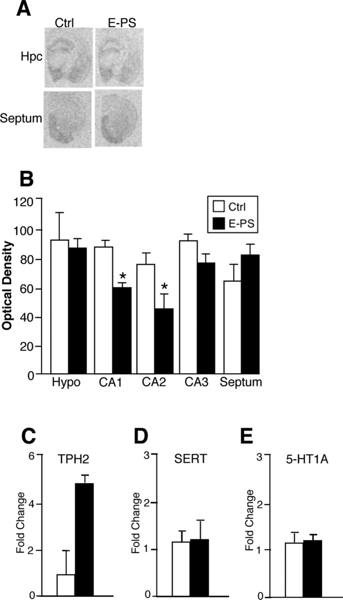
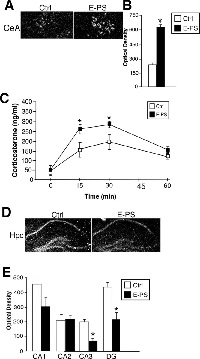
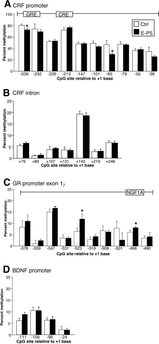
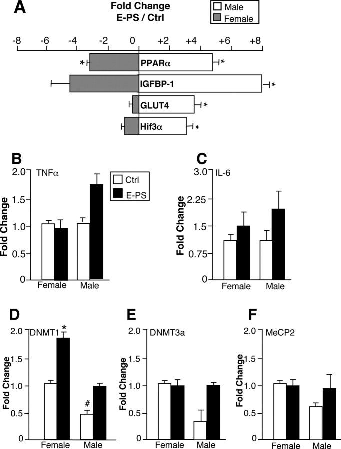
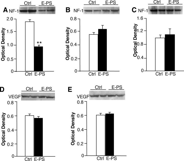
Prenatal stress is associated with an increased vulnerability to neurodevelopmental disorders, including autism and schizophrenia. To determine the critical time window when fetal antecedents may induce a disease predisposition, we examined behavioral responses in offspring exposed to stress during early, mid, and late gestation. We found that male offspring exposed to stress early in gestation displayed maladaptive behavioral stress responsivity, anhedonia, and an increased sensitivity to selective serotonin reuptake inhibitor treatment. Long-term alterations in central corticotropin-releasing factor (CRF) and glucocorticoid receptor (GR) expression, as well as increased hypothalamic-pituitary-adrenal (HPA) axis responsivity, were present in these mice and likely contributed to an elevated stress sensitivity. Changes in CRF and GR gene methylation correlated with altered gene expression, providing important evidence of epigenetic programming during early prenatal stress. In addition, we found the core mechanism underlying male vulnerability may involve sex-specific placenta responsivity, where stress early in pregnancy significantly increased expression of PPARalpha (peroxisome proliferator-activated receptor alpha), IGFBP-1 (insulin-like growth factor binding protein 1), HIF3alpha (hypoxia-inducible factor 3a), and GLUT4 (glucose transporter 4) in male placentas but not females. Examination of placental epigenetic machinery revealed basal sex differences, providing further evidence that sex-specific programming begins very early in pregnancy, and may contribute to the timing and vulnerability of the developing fetus to maternal perturbations. Overall, these results indicate that stress experience early in pregnancy may contribute to male neurodevelopmental disorders through impacts on placental function and fetal development.


Figures

Figure 1.
Figure 1.
E-PS males showed maladaptive behavioral stress responsivity in tail suspension and forced swim tests. Male and female offspring born to unstressed dams [control (C)] or dams exposed to PS during early (E), mid (M), or late (L) gestation were compared. A, B, E-PS males spent significantly more time immobile in a tail suspension test (A) and a forced swim test (B) compared with control males (*p < 0.05). C, E-PS males spent significantly less time climbing compared with control males in the forced swim test (*p < 0.05). D, No significant differences in time spent swimming were detected between control and prenatally stressed male or female offspring. E, F, No differences were found in an open field test for crosses into the center arena (E) or total number of crossed lines (F). Data are mean ± SEM. n = 4–6.
Figure 2.
Figure 2.
E-PS males showed decreased basal hedonic behavior and increased stress-induced sucrose bingeing. Anhedonia of E-PS males was examined in a sucrose preference test. A, E-PS males displayed a significant decrease in sucrose preference at a low concentration compared with Ctrl (n = 16) male offspring (*p < 0.05; n = 16). B, C, Two hours (B) and twenty-four hours (C) after an acute stress, E-PS males consumed significantly more 10% sucrose than control (*p < 0.05) and undisturbed E-PS (# p < 0.05) males after an overnight sucrose restriction (n = 8). D, E, During continuous sucrose access, an acute stress exposure did not influence 2 h (D) or 24 h (E) 10% sucrose intake for control or E-PS males (n = 4). Data are mean ± SEM.
Figure 3.
Figure 3.
E-PS males showed an increased sensitivity to acute SSRI treatment. A–C, In a tail suspension test, E-PS males spent more time immobile (A), displayed an increased number of immobility bouts (B), and showed a decreased latency to first bout of immobility (C) compared with control males (**p < 0.01). For E-PS males, a subthreshold dose of 2.5 mg/kg citalopram significantly reduced bouts of immobility (B) and latency to first bout of immobility (C; # p < 0.05) compared with vehicle. Citalopram (10 mg/kg) significantly reduced time spent immobile and bouts of immobility for control and E-PS males (# p < 0.05; ## p < 0.01) compared with vehicle. C, Citalopram (10 mg/kg) also significantly increased latency to first bout of immobility for E-PS males (# p < 0.05). Data are mean ± SEM. n = 8.
Figure 4.
Figure 4.
E-PS males showed altered serotonin transporter levels. A, B, E-PS males showed a significant reduction in SERT levels in the CA1 region of the hippocampus compared with control males (*p < 0.05). C, Real-time PCR performed on dorsal raphe micropunches revealed no significant effect of E-PS on TPH2 expression (p = 0.06). D, E, No differences were detected in SERT (D) or 5-HT1A (E) expression in the dorsal raphe. Data are mean ± SEM. n = 4–5. Hpc, Hippocampus; Hypo, hypothalamus.
Figure 5.
Figure 5.
E-PS males displayed stress pathway dysregulation including increased CeA CRF, decreased hippocampal GR, and an increased HPA axis stress response. A, B, E-PS significantly increased CRF expression in the central nucleus of the amygdala compared with controls (*p < 0.05; n = 4). C, Corticosterone levels after a 15 min restraint stress were significantly higher in E-PS males compared with controls (*p < 0.05; n = 6). D, E, GR expression was significantly lower in E-PS males compared with controls in the CA3 and DG of the dorsal hippocampus (*p < 0.05). The apparent decrease in CA1 GR expression was not significant (p = 0.07; n = 5). No effect of E-PS was seen in CA2. Data are mean ± SEM. Hpc, Hippocampus.
Figure 6.
Figure 6.
Alterations in DNA methylation of CRF and GR in E-PS males correlated with directional changes in gene expression patterns. A, Methylation analysis of the 10 CpG dinucleotides within the CRF promoter (−336 to −36 bp) revealed a significant decrease in methylation at specific cytosines in E-PS males compared with control males (*p < 0.05). B, No change in methylation was evident in the CRF intronic region (79–248 kb). C, A significant decrease in methylation at specific cytosines in E-PS males compared with control males was detected in the GR promoter region (−578 to −490 bp) (*p < 0.05). D, No changes in methylation were detected in the promoter region (−111 to −24 bp) of BDNF. Data are mean ± SEM. n = 4.
Figure 7.
Figure 7.
E-PS male placentas showed robust changes in expression of genes critical for growth and development signals. A, E-PS male placentas showed significant upregulation of PPARα, IGFBP-1, GLUT4, and HIF3α compared with male control placentas (*p < 0.01). In contrast, E-PS female placentas showed significant downregulation of PPARα (*p < 0.01) and a trending downregulation of IGFBP-1 (p = 0.06) compared with female control placentas. B, C, TNFα (B) and IL-6 (C) expression levels were not significantly altered by sex or E-PS (n = 5). D, In our analysis of sex differences in placental epigenetic machinery, control male placentas showed significantly lower expression of DNMT1 compared with control female placentas (# p < 0.01). E-PS increased expression of DNMT1 in female placentas (*p < 0.05). No effect of E-PS was detected in male DNMT1 levels. E, F, DNMT3a (E) and MeCP2 (F) expression levels were not significantly altered by sex or E-PS (n = 4). Data are mean ± SEM.
Figure 8.
Figure 8.
Significant decreases in brain NF-1 levels in E-PS males support delayed CNS development. As a gross marker of brain development, we compared NF-1 levels from postnatal day 1 male and female offspring. A, Whole-brain levels of NF-1 were significantly decreased in E-PS males compared with control male levels at postnatal day 1 (**p < 0.01). B, NF-1 levels were not different between E-PS and Ctrl females at postnatal day 1. C, Prefrontal cortex levels of NF-1 were not different between E-PS and control male adults (16 weeks). D, E, There was no significant effect of E-PS on VEGF in male (D) or female (E) postnatal day 1 brains. Data are mean ± SEM. n = 3–4.

References

    1. Altay T, Gonzales ER, Park TS, Gidday JM. Cerebrovascular inflammation after brief episodic hypoxia: modulation by neuronal and endothelial nitric oxide synthase. J Appl Physiol. 2004;96:1223–1230. discussion 1196. - PubMed
    1. Avena NM, Rada P, Moise N, Hoebel BG. Sucrose sham feeding on a binge schedule releases accumbens dopamine repeatedly and eliminates the acetylcholine satiety response. Neuroscience. 2006;139:813–820. - PubMed
    1. Bale TL, Vale WW. CRF and CRF receptors: role in stress responsivity and other behaviors. Annu Rev Pharmacol Toxicol. 2004;44:525–557. - PubMed
    1. Bale TL, Contarino A, Smith GW, Chan R, Gold LH, Sawchenko PE, Koob GF, Vale WW, Lee KF. Mice deficient for corticotropin-releasing hormone receptor-2 display anxiety-like behaviour and are hypersensitive to stress. Nat Genet. 2000;24:410–414. - PubMed
    1. Bale TL, Anderson KR, Roberts AJ, Lee KF, Nagy TR, Vale WW. Corticotropin-releasing factor receptor-2-deficient mice display abnormal homeostatic responses to challenges of increased dietary fat and cold. Endocrinology. 2003;144:2580–2587. - PubMed

Publication types

MeSH terms

Substances