

Case Reports
doi: 10.3174/ajnr.A1262.
Epub 2008 Sep 3.
Intradural hemangiopericytoma of the lumbar spine: a rare entity
Affiliations
- PMID: 18768724
- PMCID: PMC7051714
- DOI: 10.3174/ajnr.A1262
Item in Clipboard
Case Reports
Intradural hemangiopericytoma of the lumbar spine: a rare entity
AJNR Am J Neuroradiol.
2009 Jan.
Abstract
We report a case of a patient with an intradural hemangiopericytoma of the lumbar spine and the unusual MR angiography (MRA) and spinal angiography findings of arteriovenous shunting with spinal venous congestion. We highlight the concordance of the unusual MRA and angiographic findings and their relationship to combined endovascular and surgical therapy.
Figures
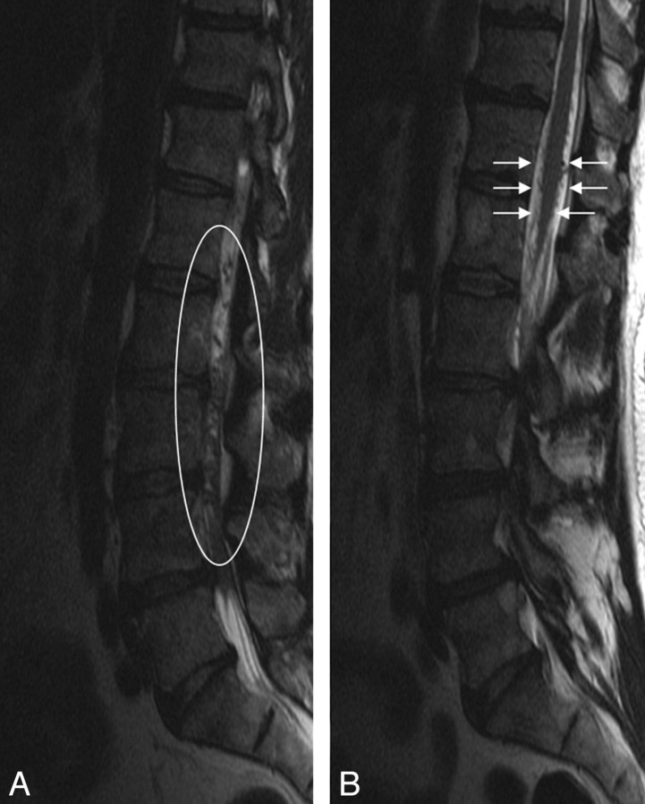
Sagittal T2-weighted images of the lumbar spine demonstrating multiple intradural flow voids (left, white oval), which appear on the surface of the conus (right, white arrows). No change in signal intensity was identified within the cord.
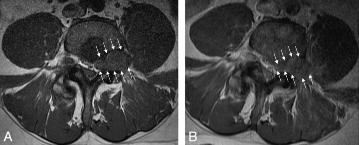
Precontrast (left) and postcontrast (right) axial T1-weighted images demonstrate an enhancing mass in the left L4 neural foramen (white arrows). The low signal intensity within the posterior aspect of the vertebral body enhanced after contrast administration, consistent with atypical hemangiomas.
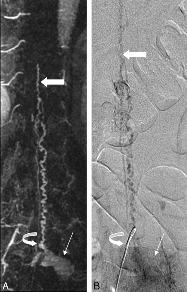
Correlative coronal contrast-enhanced spinal MRA maximum intensity projection image (left) and late-phase anteroposterior conventional spinal angiogram (right) demonstrate a hypervascular mass (thin arrow) with radicular venous drainage (curved arrow). Prominent intradural veins are present, which ascend to the level of the conus, where prominent anterior and posterior pial veins (thick arrow) are visualized.
References
-
- Harris DJ, Fornasier VL, Livingston KE. Hemangiopericytoma of the spinal canal: report of three cases. J Neurosurg 49:914–20 - PubMed
-
- Stella L, Cappabianca P, Pettinato, G, et al. Hemangiopericytoma of the meninges: report of two cases. Acta Neurologica 4:185–95 - PubMed
-
- Muraszko KM, Antunes JL, Hilal SK, et al. Hemangiopericytomas of the spine. Neurosurgery 1982;10:473–79 - PubMed
-
- Ijiri K, Yuasa S, Yone K, et al. Primary epidural hemangiopericytoma of the lumbar spine. Spine 2002;27:E189–92 - PubMed
Publication types
MeSH terms
LinkOut - more resources
Full Text Sources