. 2008 Sep 9;105(36):13556-61.
doi: 10.1073/pnas.0803055105. Epub 2008 Sep 3.

A microRNA DNA methylation signature for human cancer metastasis

Affiliations

A microRNA DNA methylation signature for human cancer metastasis

Amaia Lujambio et al. Proc Natl Acad Sci U S A. .

Abstract

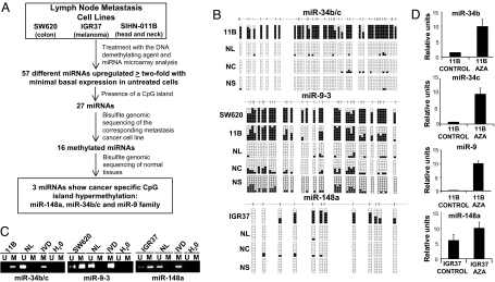
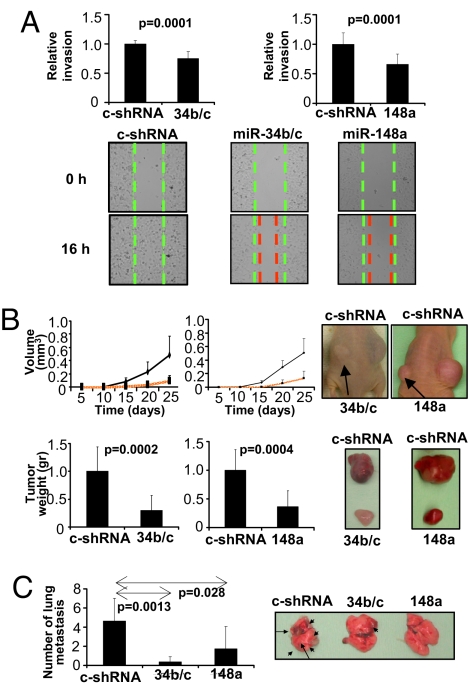
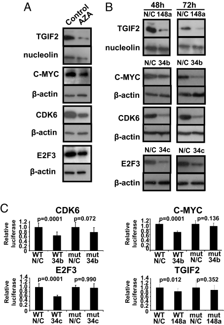
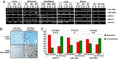
MicroRNAs (miRNAs) are small, noncoding RNAs that can contribute to cancer development and progression by acting as oncogenes or tumor suppressor genes. Recent studies have also linked different sets of miRNAs to metastasis through either the promotion or suppression of this malignant process. Interestingly, epigenetic silencing of miRNAs with tumor suppressor features by CpG island hypermethylation is also emerging as a common hallmark of human tumors. Thus, we wondered whether there was a miRNA hypermethylation profile characteristic of human metastasis. We used a pharmacological and genomic approach to reveal this aberrant epigenetic silencing program by treating lymph node metastatic cancer cells with a DNA demethylating agent followed by hybridization to an expression microarray. Among the miRNAs that were reactivated upon drug treatment, miR-148a, miR-34b/c, and miR-9 were found to undergo specific hypermethylation-associated silencing in cancer cells compared with normal tissues. The reintroduction of miR-148a and miR-34b/c in cancer cells with epigenetic inactivation inhibited their motility, reduced tumor growth, and inhibited metastasis formation in xenograft models, with an associated down-regulation of the miRNA oncogenic target genes, such as C-MYC, E2F3, CDK6, and TGIF2. Most important, the involvement of miR-148a, miR-34b/c, and miR-9 hypermethylation in metastasis formation was also suggested in human primary malignancies (n = 207) because it was significantly associated with the appearance of lymph node metastasis. Our findings indicate that DNA methylation-associated silencing of tumor suppressor miRNAs contributes to the development of human cancer metastasis.


Conflict of interest statement

The authors declare no conflict of interest.

Figures

Fig. 1.
Fig. 1.
Pharmacological epigenomic unmasking of cancer-specific hypermethylated miRNAs in metastasis cancer cells. (A) Schematic strategy used to identify DNA methylation-associated repression of miRNAs in metastatic cancer cell lines. (B) Bisulfite genomic sequencing analyses of the miR-34b/c cluster, miR-9-3, and miR-148a CpG islands in normal tissues and metastatic cell lines. Eight single clones are represented for each sample. The CpG island is depicted, and each vertical bar illustrates a single CpG. Black and white squares represent methylated and unmethylated CpG, respectively. NL, normal lymphocytes; NC, normal colon; NS, normal skin. These miRNAs show cancer-specific CpG island hypermethylation. (C) Methylation-specific PCR analyses of miR-148a, miR-34b/c, and miR-9-3. Normal lymphocytes (NL) and in vitro-methylated DNA (IVD) are shown as positive controls for the unmethylated and methylated sequences, respectively. (D) Expression analyses of mature miRNAs by qRT-PCR in methylated metastatic cell lines in untreated cells and upon treatment with the DNA demethylating agent 5-aza-2′-deoxycytidine (AZA). The use of the DNA methylation inhibitor restores miRNA expression.
Fig. 2.
Fig. 2.
miR-148a and miR-34b/c suppress tumor invasion and dissemination in vitro and in vivo. (A) Wound-healing assay. (Upper) The relative invasion of c-shRNA cells compared with miRNA transfected cells after 16 h. (Lower) Illustrative examples of the wound-healing assay in the c-shRNA and miRNA transfected metastatic cell lines at 0 h and 16 h. Green and red lines represent the initial and final leading edges of the invasion front, respectively. miR-148a and miR-34b/c transfected cells show minor invasion capability. (B) Effects of miRNA transfection on in vivo growth of metastatic SIHN-011B cells xenografted into nude mice. (Left) Tumor volume was monitored over time, and tumor weight was measured at the end of the experiment. (Right) Representative large tumors on the right flank (c-shRNA cells) and the small tumor on the opposite flank, corresponding to miR-34b/c or miR-148a transfected cells. Examples of the resected tumors are also shown. (C) Metastatic SIHN-011B cancer cells with stable transfection of miR-148a, miR-34b/c, or a control vector were transplanted into athymic nude mice by tail-vein injection. A graphical representation of the number of lung metastases observed and illustrative examples are shown.
Fig. 3.
Fig. 3.
miR-148a and miR-34b/c target and down-regulate cancer progression and metastasis-related target genes. (A) Protein expression analysis by Western blot for TGIF2, C-MYC, CDK6, and E2F3 in untreated and 5-aza-2′-deoxycytidine-treated SIHN-011B cells. The DNA demethylating agent induces down-regulation of the oncoproteins. (B) Transfection of metastatic SIHN-011B cells with the miR-148a, miR-34b, and miR-34c precursor molecules also causes a reduction in the protein levels measured. (C) Interaction between the miRNAs and the 3′ UTR of the target genes. Luciferase assays of SIHN-011B cancer cells, transfected with firefly luciferase constructs containing WT or mutant (MUT) target sites, and cotransfected with negative control (N/C) or the corresponding precursor of miRNA (miR-148a, miR-34b, or miR-34c) are shown.
Fig. 4.
Fig. 4.
The role of miRNA hypermethylation in the metastatic behavior of human primary tumors. (A) Methylation-specific PCR analyses for miR-148a, miR-34b/c, miR-9-1, miR-9-2, and miR-9-3 in primary human tumors derived from different tissues. The presence of a band under the U or M lanes indicates unmethylated or methylated sequences, respectively. Normal lymphocytes (NL) and in vitro methylated DNA (IVD) are shown as positive controls for the unmethylated and methylated sequences, respectively. (B) Association between miRNA methylation and up-regulation of their corresponding oncoprotein targets in primary tumors. Illustrative immunohistochemical examples demonstrate that C-MYC and CDK6 overexpression is associated with miR-34b/c hypermethylation in lung tumors and melanomas, respectively. (C) Graphical representation of the distribution of miR-9, miR-34b/c, and miR-148a CpG island hypermethylation in primary tumors according to the presence or absence of lymph node metastasis. The presence of hypermethylation of the miRNAs is significantly associated with the existence of metastasis in human malignancies.

Comment in

References

    1. He L, Hannon GJ. MicroRNAs: Small RNAs with a big role in gene regulation. Nat Rev Genet. 2004;5:522–531. - PubMed
    1. Bartel DP. MicroRNAs: Genomics, biogenesis, mechanism, and function. Cell. 2004;116:281–297. - PubMed
    1. Lu J, et al. MicroRNA expression profiles classify human cancers. Nature. 2005;435:834–838. - PubMed
    1. Volinia S, et al. A microRNA expression signature of human solid tumors defines cancer gene targets. Proc Natl Acad Sci USA. 2006;103:2257–2261. - PMC - PubMed
    1. Esquela-Kerscher A, Slack FJ. OncomiRs-microRNAs with a role in cancer. Nat Rev Cancer. 2006;6:259–269. - PubMed

Publication types