. 2008 Sep 15;181(6):4272-8.
doi: 10.4049/jimmunol.181.6.4272.

RAGE ligation affects T cell activation and controls T cell differentiation

Affiliations

RAGE ligation affects T cell activation and controls T cell differentiation

Yali Chen et al. J Immunol. .

Abstract

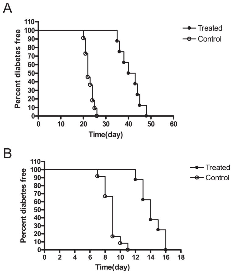
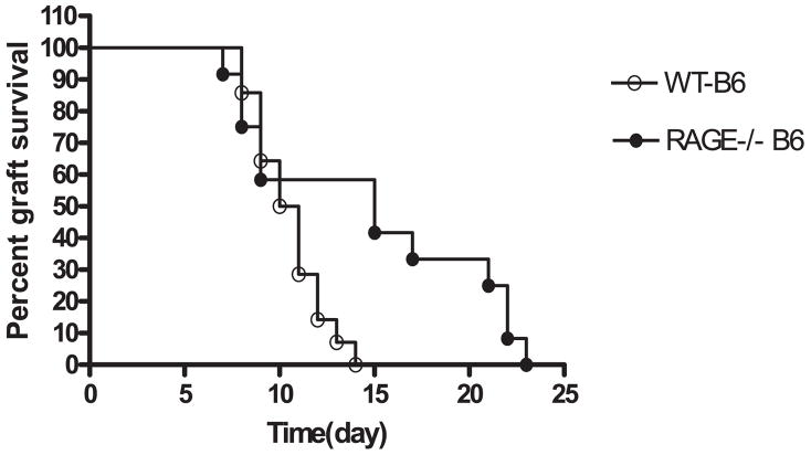
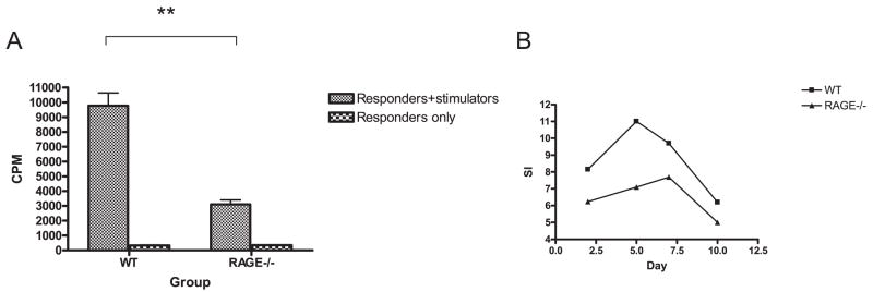
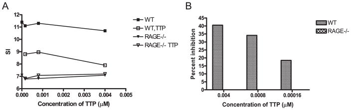
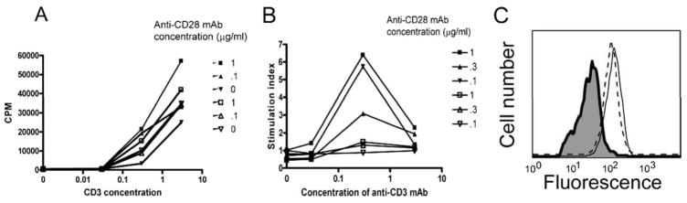
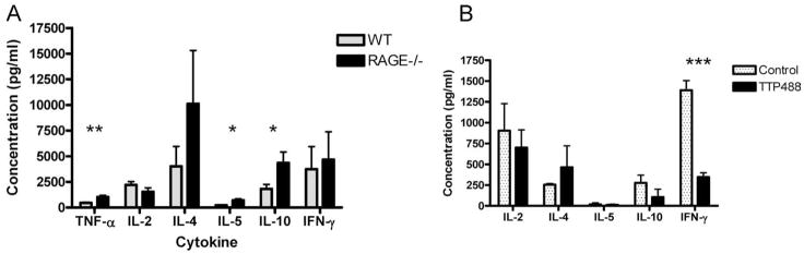
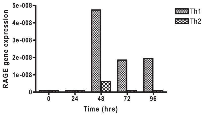
The pattern recognition receptor, RAGE, has been shown to be involved in adaptive immune responses but its role on the components of these responses is not well understood. We have studied the effects of a small molecule inhibitor of RAGE and the deletion of the receptor (RAGE-/- mice) on T cell responses involved in autoimmunity and allograft rejection. Syngeneic islet graft and islet allograft rejection was reduced in NOD and B6 mice treated with TTP488, a small molecule RAGE inhibitor (p < 0.001). RAGE-/- mice with streptozotocin-induced diabetes showed delayed rejection of islet allografts compared with wild type (WT) mice (p < 0.02). This response in vivo correlated with reduced proliferative responses of RAGE-/- T cells in MLRs and in WT T cells cultured with TTP488. Overall T cell proliferation following activation with anti-CD3 and anti-CD28 mAbs were similar in RAGE-/- and WT cells, but RAGE-/- T cells did not respond to costimulation with anti-CD28 mAb. Furthermore, culture supernatants from cultures with anti-CD3 and anti-CD28 mAbs showed higher levels of IL-10, IL-5, and TNF-alpha with RAGE-/- compared with WT T cells, and WT T cells showed reduced production of IFN-gamma in the presence of TTP488, suggesting that RAGE may be important in the differentiation of T cell subjects. Indeed, by real-time PCR, we found higher levels of RAGE mRNA expression on clonal T cells activated under Th1 differentiating conditions. We conclude that activation of RAGE on T cells is involved in early events that lead to differentiation of Th1(+) T cells.


Conflict of interest statement

Disclosures

The authors have no financial conflict of interest.

Figures

FIGURE 1
FIGURE 1
Survival of syngeneic or allogeneic islet grafts in mice with diabetes. A, NOD mice with hyperglycemia received a transplant of syngeneic islet grafts and were treated with TTP488 (n = 8) or control peptide (n = 11) as indicated in the Materials and Methods. The percentage of nondiabetic mice is shown. There was a significant prolongation of the survival of islets in mice treated with TTP488 (p < 0.001). B, C57BL/6 mice with STZ-induced diabetes were transplanted with BALB/c islets and treated with TTP488 (n = 8) or control protein (n = 8). There was a significant prolongation of graft survival with the RAGE antagonist (p < 0.001).
FIGURE 2
FIGURE 2
Survival of allogeneic islets in WT and RAGE−/− mice. Diabetes was induced with a single dose of STZ in WT (n = 14) or RAGE−/− (n = 12) B6 mice, which then received a transplant of BALB/c islets. There was a significant delay in rejection of the allogeneic (BALB/c) islets in RAGE−/− mice (p < 0.02).
FIGURE 3
FIGURE 3
Reduced MLR responses in RAGE−/− mice. A, The proliferative responses of WT or RAGE−/− T cells to BALB/c stimulators were studied in primary mixed lymphocyte reactions after 5 days. The data represent the mean values of four separate experiments. There was a significant reduction (**, p < 0.01) in the proliferative response by RAGE−/− T cells when the actual cpm or the stimulation index (cpm with stimulators/cpm without stimulators) were compared (31.0 ± 1.6 vs 9.0 ± 0.67, p < 0.001). B, The kinetics of the responses of a single experiment (representative of two) comparing WT and RAGE−/− cells are shown.
FIGURE 4
FIGURE 4
Inhibition of WT but not RAGE−/− T cells by TTP488. A, Purified WT or RAGE−/− T cells were added to irradiated BALB/c stimulator cells with TTP488 at the indicated concentrations or a similar dilution of DMSO. The incorporation of [3H]thymidine was measured after 5 days. The data are expressed as the stimulation index (cpm at the indicated concentration of TTP488 or DMSO/cpm of the responder cells only). The cpm of the responder cells were WT: 11884, RAGE−/−: 12895). B, The percent inhibition (1−cpm with TTP/cpm with equivalent dilution of DMSO *100) of the WT and RAGE−/− T cells was calculated. The data shown are from a single experiment representative of three independent experiments.
FIGURE 5
FIGURE 5
Diminished responsiveness to CD28 costimulation by RAGE−/− T cells. A, T cells were isolated from B6 (filled symbols) or RAGE−/− (open symbols) mice and added to tissue culture plates coated with the indicated concentrations of anti-CD3 mAb without or with anti-CD28 mAb at the indicated concentrations. B, The effects of CD28 costimulation were analyzed by dividing the cpm from cultures of T cells from WT or RAGE−/− T cells in the presence of the indicated concentrations of anti-CD28 mAb by the cpm from cultures without CD28. There was markedly reduced response to CD28 costimulation by the RAGE−/− T cells. The data shown is from a single experiment that is representative of three independent studies. C, To exclude differences in CD28 expression as the basis for the findings in B, the expression of CD28 was analyzed on WT (dashed line) and RAGE−/− (solid line) CD4+ T cells by flow cytometry (the isotype control is shaded).
FIGURE 6
FIGURE 6
Cytokine concentrations in the supernatants of activated T cells. A, Purified RAGE−/− (solid bars) and WT (stippled bars) T cells were activated with anti-CD3 and anti-CD28 mAbs and the culture supernatants were harvested for measurement of cytokine concentrations (mean ± SEM, n = 4/group). There was a significant increase in the concentration of TNF-α, IL-5, and IL-10 in the cultures from RAGE−/− mice compared with WT mice (*, p < 0.05; **, p < 0.01). B, Primary MLR’s with purified WT T cells were cultured in the presence of TTP488 or the excipient DMSO as described. The responders were harvested after 5 days and activated with PMA/ionomycin overnight. The concentrations in the supernatants were measured (n = 3/group). There was a significant reduction in the production of IFN-γ in responders that had been cultured with TTP488.
FIGURE 7
FIGURE 7
Expression of RAGE transcripts in AND transgenic T cells cultured under Th1 and Th2 differentiating conditions. RAGE transcripts were measured by real-time PCR and compared with actin in naive AND transgenic T cells cultured with mitomycin C-treated APC’s and Ag under Th1 (fine hatched bar) and Th2 (coarse hatched bar) skewing conditions as described in Materials and Methods. The data are from a single experiment that is representative of two independent experiments.

References

    1. Zanin-Zhorov A, Bruck R, Tal G, Oren S, Aeed H, Hershkoviz R, Cohen IR, Lider O. Heat shock protein 60 inhibits Th1-mediated hepatitis model via innate regulation of Th1/Th2 transcription factors and cytokines. J Immunol. 2005;174:3227–3236. - PubMed
    1. Alyanakian MA, Grela F, Aumeunier A, Chiavaroli C, Gouarin C, Bardel E, Normier G, Chatenoud L, Thieblemont N, Bach JF. Transforming growth factor-β and natural killer T-cells are involved in the protective effect of a bacterial extract on type 1 diabetes. Diabetes. 2006;55:179–185. - PubMed
    1. Zipris D, Lien E, Nair A, Xie JX, Greiner DL, Mordes JP, Rossini AA. TLR9-signaling pathways are involved in Kilham rat virus-induced autoimmune diabetes in the biobreeding diabetes-resistant rat. J Immunol. 2007;178:693–701. - PubMed
    1. Thomas HE, Irawaty W, Darwiche R, Brodnicki TC, Santamaria P, Allison J, Kay TW. IL-1 receptor deficiency slows progression to diabetes in the NOD mouse. Diabetes. 2004;53:113–121. - PubMed
    1. Sandberg JO, Eizirik DL, Sandler S. IL-1 receptor antagonist inhibits recurrence of disease after syngeneic pancreatic islet transplantation to spontaneously diabetic non-obese diabetic (NOD) mice. Clin Exp Immunol. 1997;108:314–317. - PMC - PubMed

Publication types

MeSH terms

Substances