. 2008 Nov;82(22):11045-53.
doi: 10.1128/JVI.01593-08. Epub 2008 Sep 3.

Novel characteristics of the function and induction of murine p56 family proteins

Affiliations

Novel characteristics of the function and induction of murine p56 family proteins

Volker Fensterl et al. J Virol. 2008 Nov.

Abstract

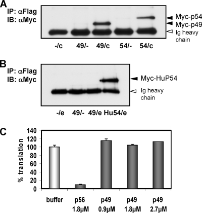
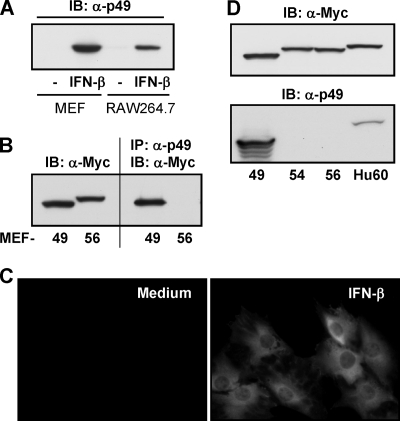
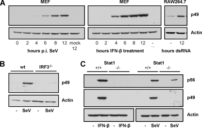
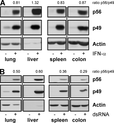
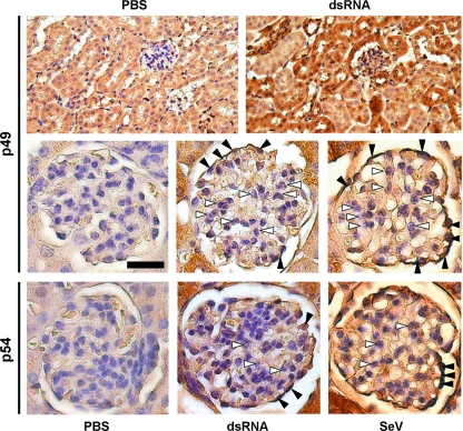
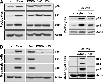
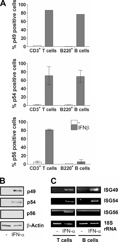
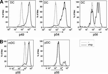
The interferon-stimulated gene 56 (ISG56) family is induced strongly in response to virus infection, interferons (IFNs) and double-stranded RNA (dsRNA). In the mouse, this family comprises three members, ISG56, ISG54, and ISG49, which are clustered on chromosome 19 and encode the corresponding proteins p56, p54, and p49. Here, we report differential properties of these proteins and their distinct induction patterns in different cell types. All three murine proteins bound to the c-subunit of the translation initiation factor eIF3, but unlike the other members, p49 did not inhibit protein synthesis. Using a newly raised antibody, we demonstrated that both in vitro and in vivo, p49 expression was strongly induced by IFN, dsRNA, and Sendai virus. However, in kidney mesangial cells, as opposed to podocytes, encephalomyocarditis virus, vesicular stomatitis virus, or extracellular dsRNA did not induce any of the p56 family proteins, although they were robustly expressed after Sendai virus infection or dsRNA transfection. Furthermore, protein-specific differences in the regulation of p56 family members became evident in various leukocyte types: all three proteins were induced by IFN in T cells, but in B cells p56 and ISG56 mRNA could not be detected. Similarly, p56 was selectively uninducible in plasmacytoid dendritic cells, whereas in myeloid dendritic cells, all three family members were expressed. These results revealed novel cell type-, inducer-, and gene-specific regulation of the ISG56 family of genes.


Figures

FIG. 1.
FIG. 1.
p49 binds to eIF3c but does not inhibit translation. (A) HT1080 cells were cotransfected with expression vectors for Myc-tagged murine p49 or murine p54 and Flag-tagged eIF3c. After Flag-immunoprecipitation, coprecipitated Myc-tagged proteins were detected by immunoblotting. (B) HT1080 cells were cotransfected with expression vectors for Myc-tagged murine p49 or human p54 and Flag-tagged eIF3e. After Flag immunoprecipitation, coprecipitated Myc-tagged proteins were detected by immunoblotting. (C) Translation inhibition by p49 and p56 was quantified in vitro by using reticulocyte lysate-driven luciferase mRNA translation in the presence of [35S]cysteine. Densitometric averages of polyacrylamide gel electrophoresis-resolved luciferase signal are shown.
FIG. 2.
FIG. 2.
Specificity of p49 antibody and cellular localization of p49. (A) The newly raised p49 antibody was used to detect endogenous p49 by immunoblotting after 18 h treatment of MEFs or RAW 264.7 cells with 1,000 U of IFN-β/ml. (B) Immunoprecipitation of p49 was done with cell lysates of MEFs stably expressing Myc-p49 or Myc-p56. Precipitated protein was detected by Myc-immunoblotting. (C) Specificity of p49 antibody was demonstrated with cell lysates from HT1080 cells transfected with vectors for expression of Myc-p49, -p54, or -p56 or Myc-human P60, the homolog of murine p49. (D) Cellular localization of endogenous p49, induced in MEFs by 16 h of treatment with IFN-β, was examined by immunofluorescence.
FIG. 3.
FIG. 3.
Induction of p49 by SeV infection requires IFN. (A) p49 expression was detected by immunoblotting of cell lysates of either MEFs, after infection with 80 hemagglutinating units (HAU) of SeV/ml or treatment with 1,000 U of IFN-β/ml for the indicated times, or of RAW 264.7 cells, after the addition of dsRNA [100 μg of poly(IC)/ml] to the medium for 12 h. (B) IRF-3 dependence of virus-induced p49 expression was demonstrated by infecting wild-type or IRF3−/− MEFs with 80 HAU of SeV/ml for 16 h and immunoblotting cell lysates, detecting protein expression with p49 or actin antibodies. (C) IFN dependence of virus-induced p49 but not p56 expression was demonstrated by treating wild-type or Stat1−/− MEFs with IFN-β or infecting the cells with 80 HAU of SeV/ml for 16 h and immunoblotting the cell lysates.
FIG. 4.
FIG. 4.
Induction of p49 and p56 in mice injected with IFN or dsRNA. At 8 h after i.v. injection of IFN-α (A) or dsRNA (B) or PBS, mice were sacrificed and organs were homogenized. Organ extracts (40 μg in panel A and 20 μg in panel B) were examined for expression of p49, p56, or actin by immunoblotting.
FIG. 5.
FIG. 5.
Induction of p49 and p54 in the glomeruli of murine kidneys. At 8 h after i.v. injection of dsRNA, SeV, or PBS, mice were sacrificed, kidneys were collected, fixed, and processed for immunohistochemistry to detect p49 (upper and middle panel) and p54 (lower panel). White arrowheads indicate mesangial cells, black arrowheads indicate podocytes in the blood-filtrating glomeruli, located in the renal cortex. Magnifications are ×200 (upper panel) and ×400 (middle and lower panels). Scale bar, 25 μm.
FIG. 6.
FIG. 6.
Selective inducibility of p56 family members in mesangial cells. Differentiated podocytes (A) and mesangial cells (B) were either treated with 500 U of IFN-α/ml or infected at a multiplicity of infection of 10 with EMCV, SeV, or VSV for 8 h. Expression of p49, p54, and p56 was detected by immunoblotting. Alternatively, cells were exposed to dsRNA [poly(IC)] by either FuGENE 6-mediated transfection (cytopl.) or by adding poly(IC) to the medium (float). In mesangial cells, infection by VSV was verified by detection of P-protein and EMCV infection by observation of development of the cytopathic effect (not shown).
FIG. 7.
FIG. 7.
Lack of induction of p56 by IFN in B cells in vitro. (A) Peripheral blood cells were isolated from mice and stimulated with 1,000 U of IFN-β/ml (106 cells/sample). After stimulation (24 h), cells were harvested and stained for flow cytometry. (B) Murine B cells isolated from spleens were untreated or treated with IFN-α for 24 h, and extracts were immunoblotted for the indicated proteins. (C) After 4 h of stimulation of isolated B or T cells with 1,000 U of IFN-α/ml, RNA was isolated. cDNA produced from this RNA provided the template for PCR using primers for ISG49, ISG54, ISG56, and 18S rRNA.
FIG. 8.
FIG. 8.
p56 is induced in mDCs but not pDCs. (A) Dendritic cells were differentiated from mouse bone marrow using granulocyte-macrophage colony-stimulating factor. Cells were stimulated for 16 h with 1,000 U of IFN-β/ml before analysis by flow cytometry using antibodies to p49, p54, and p56. (B) Dendritic cells differentiated from bone marrow using Flt3-ligand were stimulated for 16 h with 1,000 U of IFN-β/ml before analysis by flow cytometry. pDC and mDC populations were distinguished by using surface markers, and the p56 expression was detected.

References

    1. Alexopoulou, L., A. C. Holt, R. Medzhitov, and R. A. Flavell. 2001. Recognition of double-stranded RNA and activation of NF-κB by Toll-like receptor 3. Nature 413732-738. - PubMed
    1. Andersen, J., S. VanScoy, T. F. Cheng, D. Gomez, and N. C. Reich. 2008. IRF-3-dependent and augmented target genes during viral infection. Genes Immun. 9168-175. - PubMed
    1. Andrejeva, J., K. S. Childs, D. F. Young, T. S. Carlos, N. Stock, S. Goodbourn, and R. E. Randall. 2004. The V proteins of paramyxoviruses bind the IFN-inducible RNA helicase, mda-5, and inhibit its activation of the IFN-beta promoter. Proc. Natl. Acad. Sci. USA 10117264-17269. - PMC - PubMed
    1. Bluyssen, H. A., R. J. Vlietstra, P. W. Faber, E. M. Smit, A. Hagemeijer, and J. Trapman. 1994. Structure, chromosome localization, and regulation of expression of the interferon-regulated mouse Ifi54/Ifi56 gene family. Genomics 24137-148. - PubMed
    1. D'Andrea, L. D., and L. Regan. 2003. TPR proteins: the versatile helix. Trends Biochem. Sci. 28655-662. - PubMed

Publication types

MeSH terms

LinkOut - more resources