

Peritubular myoid cells are not the migrating population required for testis cord formation in the XY gonad
- PMID: 18769072
- PMCID: PMC2683975
- DOI: 10.1159/000143430
Peritubular myoid cells are not the migrating population required for testis cord formation in the XY gonad
Abstract
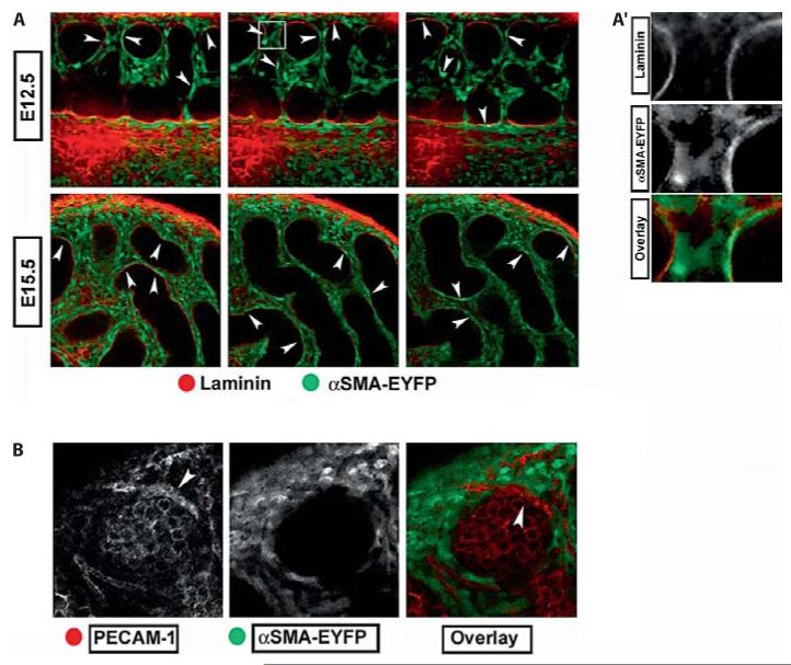
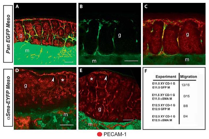
Cell migration is one of the earliest events required for development of the testis. Migration occurs only in XY gonads downstream of Sry expression and is required for the subsequent epithelialization of testis cords. Using organ culture experiments and tissue recombination, we and others speculated that peritubular myoid (PTM) cells were among the migratory cells and were likely the cell type required for cord formation. However, because no unique marker was found for PTM cells, their positive identification during or after migration remained unclear. alpha-Smooth Muscle Actin (alphaSma; approved gene symbol Acta2), a classic marker of adult PTM cells,is expressed broadly in testis interstitial cells at E12.5, and becomes highly enriched in PTM cells by E15.5-16.5. We used a novel transgenic line expressingEYFP under the control of an alphaSma promoter to determine whether alphaSma-EYFP positive cellsmigrate into the gonad. Surprisingly, mesonephroi expressing alphaSma-EYFP do not contribute any EYFP positive cells to XY gonads when used as donors in recombination cultures. These results indicate that alphaSma-EYFP cells do not migrate into the gonad during the critical window of sex determination and cannot be the migrating cell type required for testis cord formation. Our results suggest that PTM cells, and most other interstitial lineages, with the exception of endothelial cells, are induced within the gonad. These experiments suggest that endothelial cells are the migrating cell type required for epithelialization of testis cords.
Copyright 2008 S. Karger AG, Basel.
Figures
References
-
- Brennan J, Karl J, Capel B. Divergent vascular mechanisms downstream of Sry establish the arterial system in the XY gonad. Dev Biol. 2002;244:418–428. - PubMed
-
- Buehr M, Gu S, McLaren A. Mesonephric contribution to testis differentiation in the fetal mouse. Development. 1993;117:273–281. - PubMed
-
- Jeanes A, Wilhelm D, Wilson MJ, Bowles J, McClive PJ, et al. Evaluation of candidate markers for the peritubular myoid cell lineage in the developing mouse testis. Reproduction. 2005;130:509–516. - PubMed
-
- Lammert E, Cleaver O, Melton D. Induction of pancreatic differentiation by signals from blood vessels. Science. 2001;294:564–567. - PubMed
MeSH terms
Substances
Grants and funding
LinkOut - more resources
Full Text Sources
Miscellaneous
