. 2008 Sep 15;7(18):2795-802.
doi: 10.4161/cc.7.18.6687. Epub 2008 Sep 28.

The yin and yang of the Cdkn2a locus in senescence and aging

Affiliations

The yin and yang of the Cdkn2a locus in senescence and aging

Darren J Baker et al. Cell Cycle. .

Abstract

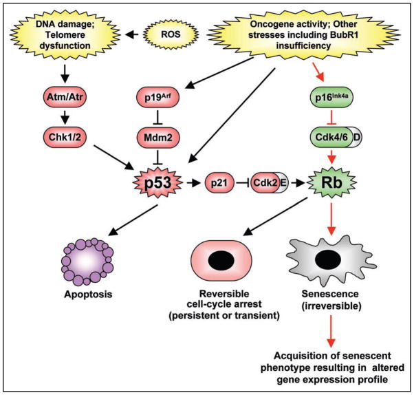
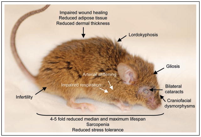
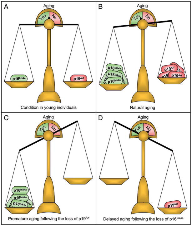
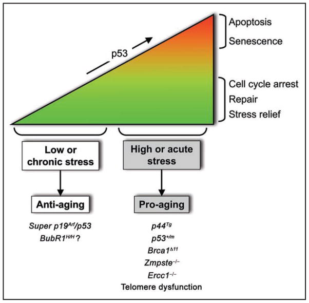
Senescence of cultured cells involves activation of the p19(Arf)-p53 and the p16(Ink4a)-Rb tumor suppressor pathways. This, together with the observation that p19(Arf) and p16(Ink4a) expression increases with age in many tissues of humans and rodents, led to the speculation that these pathways drive in vivo senescence and natural aging. However, it has been difficult to test this hypothesis using a mammalian model system because inactivation of either of these pathways results in early death from tumors. One approach to bypass this problem would be to inactivate these pathways in a murine segmental progeria model such as mice that express low amounts of the mitotic checkpoint protein BubR1 (BubR1 hypomorphic mice). These mice have a five-fold reduced lifespan and develop a variety of early-aging associated phenotypes including cachetic dwarfism, skeletal muscle degeneration, cataracts, arterial stiffening, (subcutaneous) fat loss, reduced stress tolerance and impaired wound healing. Importantly, BubR1 hypomorphism elevates both p16(Ink4a) and p19(Arf) expression in skeletal muscle and fat. Inactivation of p16(Ink4a) in BubR1 mutant mice delays both cellular senescence and aging specifically in these tissues. Surprisingly, however, inactivation of p19(Arf) has the opposite effect; it exacerbates in vivo senescence and aging in skeletal muscle and fat. These mouse studies suggest that p16(Ink4a) is indeed an effector of aging and in vivo senescence, but p19(Arf) an attenuator. Thus, the role of the p19(Arf)-p53 pathway in aging and in vivo senescence seems far more complex than previously anticipated.


Figures

Figure 1
Figure 1
Senescence controlled by the p16Ink4a-Rb and p19Arf-p53 pathways. Various intrinsic and extrinsic stress-inducing signals are able to engage the tumor suppressor pathways containing p53, p16Ink4a-Rb or both. Activation of p53 triggers cell death by inducing expression of a number of downstream target genes implicated in apoptosis (e.g., Bim, Puma, Noxa, Bid, Bax and Apaf1) or a transient or permanent (though reversible) cell cycle arrest via induction of p21. On the other hand, Rb is capable of inducing an irreversible state of cellular senescence via repression of E2F target genes and alterations in chromatin structure.
Figure 2
Figure 2
Summary of tissues affected with age-related pathologies in BubR1 hypomorphic mice. Shown is a 5-month-old BubR1 insufficient animal. Highlighted are several of the progeroid features commonly observed in this model.
Figure 3
Figure 3
The Cdkn2a locus and aging. (A) In young mice, the amount of p16Ink4a and p19Arf is quite low and in balance. (B) As animals age, p16Ink4a and p19Arf accumulate in the same tissue, with p19Arf attempting to counter-balance the pro-aging effects of p16Ink4a. With time, the balance shifts towards a state of aging. (C) In scenarios where p19Arf is not present to counteract p16Ink4a, the absolute levels of p16Ink4a increase dramatically, further promoting a pro-aging microenvironment. (D) When p16Ink4a is absent, p19Arf does not accumulate. Aging is delayed and the induction of p19Arf is not required.
Figure 4
Figure 4
Working model for regulation of aging by the p19Arf-p53 pathway. Various stresses engage and stabilize p53. Acute situations mobilize an intense p53 response, leading to apoptosis or cellular senescence and the establishment of early aging. Chronic/low amounts of stress on the other hand, might activate p53 to provide a transient cell cycle arrest that provides an opportunity to repair damage or relieve cellular stress, thereby improving cell function in the presence of stress. Stress encountered with natural aging might engage a response similar to that of chronic stress.
Figure 5
Figure 5
BubR1 deficient cells have normal DNA damage responses. (A) Passage 5 mouse embryonic fibroblasts (MEFs) of the indicated genotypes were treated with doxorubicin (Dox) for the indicated times as described. Cell lysates were generated and western blotting was performed as described. Blots were probed for BubR1, Parp-1 and γH2AX. Position of p89, the major degradation product of Parp-1 is indicated. Antibodies for BubR1, Parp-1, and γH2AX were as described. Actin was used as a loading control. These results suggest an intact response to doxorubicin-induced damage in MEFs expressing low amounts of BubR1. (B) Additional DNA damage-induced cell death analysis in BubR1 hypomorphic cells. Passage 5 MEFs were treated with actinomycin D (Act D), or a combination of Act D and anti-Fas or TNFα and compared to non-treated MEFs as described. These blots suggest that signals that activate apoptosis are interpreted similarly in wildtype and BubR1 hypomorphic MEFs. (C) Comet assay results from 5-month-old tissues of wildtype and BubR1 hypomorphic mice. Comet assays were performed according to manufacturer’s protocol (Trevigen). In this assay, both the percentage of nuclei demonstrating DNA damage as well as the amount of damage reflected by tail length, are equal irrespective of genotype, suggesting that BubR1 hypomorphic mice exhibit a normal DNA damage response to in vivo stresses. γ-irradiation was used as a positive control in the Comet assays.

Similar articles

Cited by

References

    1. Collado M, Blasco MA, Serrano M. Cellular senescence in cancer and aging. Cell. 2007;130:223–33. - PubMed
    1. Campisi J, d’Adda di Fagagna F. Cellular senescence: when bad things happen to good cells. Nat Rev Mol Cell Biol. 2007;8:729–40. - PubMed
    1. Kamijo T, Zindy F, Roussel MF, Quelle DE, Downing JR, Ashmun RA, Grosveld G, Sherr CJ. Tumor suppression at the mouse INK4a locus mediated by the alternative reading frame product p19ARF. Cell. 1997;91:649–59. - PubMed
    1. Sherr CJ. The INK4a/ARF network in tumour suppression. Nat Rev Mol Cell Biol. 2001;2:731–7. - PubMed
    1. Campisi J. Senescent cells, tumor suppression and organismal aging: good citizens, bad neighbors. Cell. 2005;120:513–22. - PubMed

Publication types

Substances