. 2008 Sep 4;3(9):e3135.
doi: 10.1371/journal.pone.0003135.

Mechanisms of hybrid oligomer formation in the pathogenesis of combined Alzheimer's and Parkinson's diseases

Affiliations

Mechanisms of hybrid oligomer formation in the pathogenesis of combined Alzheimer's and Parkinson's diseases

Igor F Tsigelny et al. PLoS One. .

Retraction in

Abstract

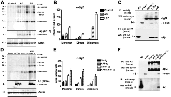
Background: Misfolding and pathological aggregation of neuronal proteins has been proposed to play a critical role in the pathogenesis of neurodegenerative disorders. Alzheimer's disease (AD) and Parkinson's disease (PD) are frequent neurodegenerative diseases of the aging population. While progressive accumulation of amyloid beta protein (Abeta) oligomers has been identified as one of the central toxic events in AD, accumulation of alpha-synuclein (alpha-syn) resulting in the formation of oligomers and protofibrils has been linked to PD and Lewy body Disease (LBD). We have recently shown that Abeta promotes alpha-syn aggregation and toxic conversion in vivo, suggesting that abnormal interactions between misfolded proteins might contribute to disease pathogenesis. However the molecular characteristics and consequences of these interactions are not completely clear.

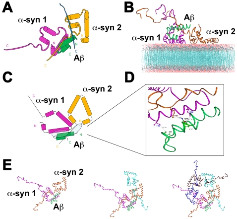
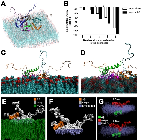
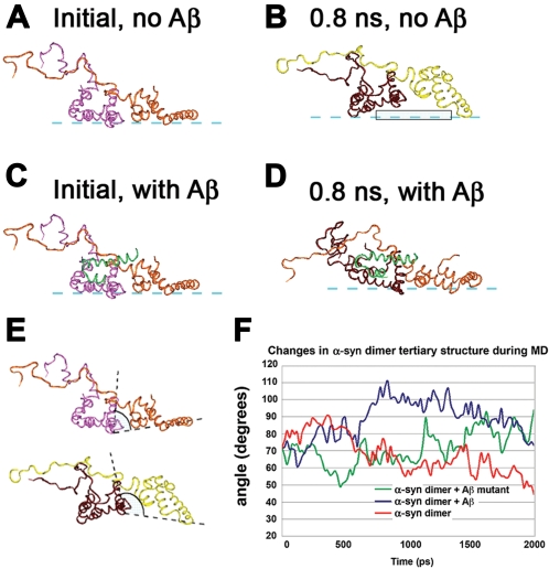
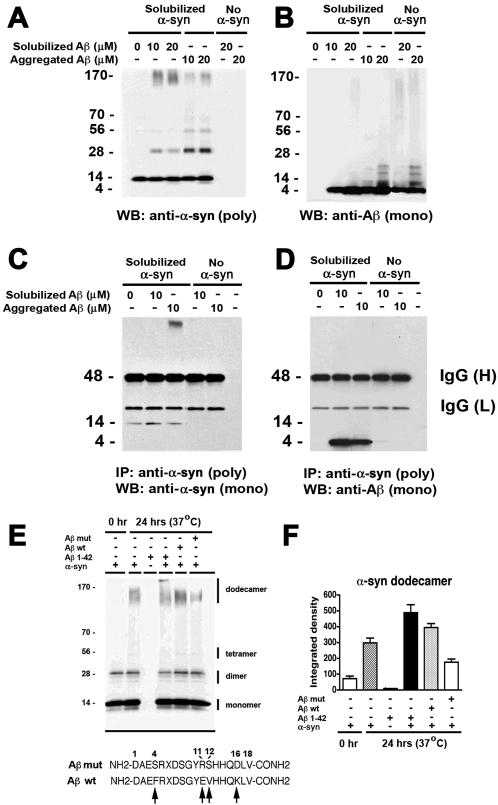
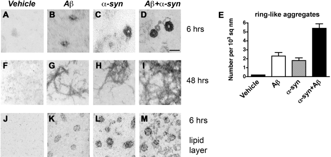
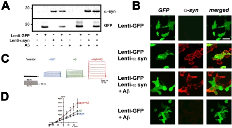
Methodology/principal findings: In order to understand the molecular mechanisms involved in potential Abeta/alpha-syn interactions, immunoblot, molecular modeling, and in vitro studies with alpha-syn and Abeta were performed. We showed in vivo in the brains of patients with AD/PD and in transgenic mice, Abeta and alpha-synuclein co-immunoprecipitate and form complexes. Molecular modeling and simulations showed that Abeta binds alpha-syn monomers, homodimers, and trimers, forming hybrid ring-like pentamers. Interactions occurred between the N-terminus of Abeta and the N-terminus and C-terminus of alpha-syn. Interacting alpha-syn and Abeta dimers that dock on the membrane incorporated additional alpha-syn molecules, leading to the formation of more stable pentamers and hexamers that adopt a ring-like structure. Consistent with the simulations, under in vitro cell-free conditions, Abeta interacted with alpha-syn, forming hybrid pore-like oligomers. Moreover, cells expressing alpha-syn and treated with Abeta displayed increased current amplitudes and calcium influx consistent with the formation of cation channels.

Conclusion/significance: These results support the contention that Abeta directly interacts with alpha-syn and stabilized the formation of hybrid nanopores that alter neuronal activity and might contribute to the mechanisms of neurodegeneration in AD and PD. The broader implications of such hybrid interactions might be important to the pathogenesis of other disorders of protein misfolding.


Conflict of interest statement

Competing Interests: The authors have declared that no competing interests exist.

Figures

Figure 1
Figure 1. Immunoblot and co-immunoprecipitation analysis of aggregated α-syn in brains of LBD cases and tg mice.
(A, B) Representative western blot (A) and semi-quantitative analysis (B) of levels of α-syn dimers and oligomers in membrane fractions from the frontal cortex of age-matched non-demented control, AD and LBD brains. In cases with LBD there was a significant increase in the levels of α-syn dimers and oligomers when compared to controls. (C) Immunoprecipitation of homogenates from control, LBD and AD cases with monoclonal antibodies against Aβ (upper panel) or α-syn (lower panel). Immunoblots were probed with monoclonal antibodies against α-syn (upper panel) or Aβ (lower panel). Aβ and α-syn pure proteins were included on the immunoblots as positive controls (first and second lanes). (D, E) Representative western blot (D) and semi-quantitative analysis (E) of levels of α-syn dimers and oligomers in the membrane fractions from nontg, APP tg, α-syn tg, and APP/α-syn double tg brains. In APP/α-syn tg mice there was a significant increase in the levels of α-syn dimers and oligomers when compared to nontg or single tg animals. (F) Immunoprecipitation of brain homogenates from nontg, APP tg, α-syn tg, and APP/α-syn double tg animals with monoclonal antibodies against Aβ (upper panel) or α-syn (lower panel). Immunoblots were probed with monoclonal antibodies against α-syn (upper panel) or Aβ (lower panel). Bar graphs represent the mean of n = 4 cases per group. *P<0.05 compared to control human brains or nontg mouse brains (by one-way ANOVA with post-hoc Tukey-Kramer test).
Figure 2
Figure 2. Molecular dynamics studies of the interactions of one Aβ monomer with membrane-docked α-syn multimers.
(A) Diagrammatic representation of the complex formed between the 2 ns Aβ conformer and the 4 ns α-syn dimer, composed of two molecules of α-syn (α-syn1 and α-syn2). (B) Molecular modeling of the Aβ monomer/α-syn dimer complex docked to the membrane. (C) Diagrammatic representation of predicted interactions between the Aβ monomer and the two α-syn molecules occur primarily between the N-terminus of Aβ and the N-terminus of the α-syn molecules (circled regions). (D) Specific residues involved in interactions between the Aβ monomer and the two α-syn molecules. (E) Docked complex of Aβ and the α-syn dimer or higher-order oligomers (trimer, pentamer) form a ring-like structure on the membrane.
Figure 3
Figure 3. Modeling and energies of interaction between Aβ and α-syn oligomers docked on the membrane.
(A) Conformation of lowest-energy complex between α-syn pentamer and 2 ns Aβ conformer on the membrane. (B) Calculated values for the most favorable energies of interaction between one Aβ monomer and an α-syn monomer, dimer, trimer, tetramer or pentamer. Compared to α-syn homomeric species composed of the same number of α-syn molecules, hybrid Aβ/α-syn multimers were more stable and had more favorable (lower) electrostatic energies of interaction. (C, D) Hybrid Aβ/α-syn multimers formed ring-like structures that embedded in the membrane after 350–800 ps of simulation. (E) Space-filled model of Aβ (orange)/α-syn (gray) multimer embedded in the membrane (green) at a depth of 1.6Å after 800 ps of MD simulation. (F) Space-filled model of Aβ (orange)/α-syn (gray) multimer showing the depth of 1.6Å (purple) that the complex embedded into the membrane (POPC) after 800 ps of MD simulation. (G) Space-filled model of Aβ (green) and α-syn (red) monomer initially situated on opposite sides of the POPC membrane showing penetration of the Aβ and α-syn molecules into the membrane and interaction between the two after 2.3 ns of MD simulation.
Figure 4
Figure 4. Molecular dynamics of conformational changes of membrane-associated α-syn in the presence of one Aβ monomer.
(A) Initial conformation of α-syn dimer on the membrane without Aβ. (B) 0.8 ns conformation of α-syn dimer on the membrane without Aβ. (C) Initial conformation of α-syn dimer on the membrane with Aβ monomer. (D) When complexed with the Aβ monomer, after 0.8 ns the conformation of the α-syn dimer on the membrane is drawn closer to the membrane, and the α-syn molecules make more contact points with the membrane surface than the α-syn dimer alone. (E) Comparison of the two complexes at 0.8 ns demonstrating the angle measured between the C-alpha atoms of the residues VAL66 (α-syn1), LYS45 (α-syn1), VAL37 (α-syn2). The complex without Aβ had an angle of <80° (upper), while the complex with Aβ had an angle of >80° (lower) and was more stable on the membrane. (F) Changes in angle measurements in the complexes over time without Aβ or in the presence of wild-type or mutated full-length Aβ. In the mutated Aβ peptide, positively-charged residues were substituted for negative ones and hydrophobic residues for hydrophilic ones (PHE4SER, GLU11ARG, VAL12SER, LYS16ASP).
Figure 5
Figure 5. Interactions between Aβ and α-syn in an in vitro cell-free aggregation model.
Freshly-solubilized or pre-aggregated Aβ1–42 was incubated at concentrations of 10 or 20 µM (as indicated) with freshly-solubilized recombinant α-syn and samples were analyzed by immunoblot or co-immunoprecipitation. (A) Increased levels of α-syn high-molecular weight forms aggregates in the presence of freshly-solubilized or pre-aggregated Aβ, as shown by immunoblot analysis with a polyclonal antibody against α-syn. (B) Formation of Aβ multimers after incubation under aggregating conditions. (C, D) In vitro binding assay for Aβ and α-syn. Immunoprecipitation of cell-free aggregated samples was performed using a polyclonal antibody against α-syn. Immunoblots were probed with monoclonal antibodies against α-syn (C) or Aβ (D). Aβ was only detected in samples that were incubated with α-syn, and in cases where no recombinant protein was included (negative control), no immunoreactivity was detected with either antibody. (E, F) Samples containing freshly-solubilized α-syn were incubated with the following peptides: an 18-mer N-terminal fragment of Aβ containing mutations at the residues determined to be critical for interaction with α-syn (Aβ mut), an 18-mer N-terminal fragment of Aβ of the wild-type sequence (Aβ wt), or full-length Aβ1–42 peptide (Aβ 1–42) and analyzed by immunoblot with an antibody against α-syn. Incubation with the Aβ mut resulted in lower levels of the dodecamer form of α-syn. The sequence of the wt and mut Aβ peptides (18-mers) used for these experiments are shown in panel e. Arrows indicate mutated residues.
Figure 6
Figure 6. Electron microscopy analysis of hybrid oligomers and fibrils of Aβ and α-syn.
In vitro cell-free preparations of vehicle, Aβ alone, α-syn alone, or Aβ and α-syn were incubated for 6 hrs or 48 hrs and prepared for electron microscopy analysis. (A–D) After 6 hrs of incubation, compared to vehicle alone (A), Aβ (B) and α-syn (C) alone formed globular structures of 5–10 nm in diameter, while incubation of Aβ and α-syn together (D) resulted in the formation of larger, well-defined ring-like structures with a central channel. (E) Analysis of numbers of ring-like structures formed after 6 hrs incubation. (F–I) After 48 hrs of incubation, compared to vehicle alone (F), Aβ (G) and α-syn (H) alone or in combination (I) formed fibrils of about 11 nm in diameter. (J–M) After 6 hrs incubation in the presence of lipid monolayers, compared to vehicle alone (J), Aβ (K) and α-syn (L) alone formed globular structures of 5–8 nm in diameter, while incubation of Aβ and α-syn together (M) resulted in enhanced formation of larger, well-defined, ring-like structures. Scale bar, 10 nm (A–D, J–M); 100 nm (F–I).
Figure 7
Figure 7. Electrophysiological analysis of the cellular effects of hybrid Aβ/α-syn multimers.
293T cells were infected with lenti-α-syn and lenti-GFP and treated with Aβ and analyzed by electrophysiology. (A) Immunoblot analysis showing expression levels of α-syn and GFP in treated cultures. (B) Immunocytochemical analysis demonstrating high efficiency of infection and co-localization between α-syn and GFP. (C, D) Representative currents (C) elicited by depolarizing the cells from a holding potential of −50 mV to a series of test potentials ranging from −80 mV to +80 mV, and corresponding current voltage-relationship curves (D), in α-syn-expressing cells treated with vehicle (α-syn, n = 5), vector-transfected cells treated with Aβ (Aβ, n = 5), and α-syn-expressing cells treated with Aβ (α-syn+Aβ, n = 6).

References

    1. Lashuel HA, Hartley DM, Petre BM, Wall JS, Simon MN, et al. Mixtures of wild-type and a pathogenic (E22G) form of Abeta40 in vitro accumulate protofibrils, including amyloid pores. J Mol Biol. 2003;332:795–808. - PubMed
    1. Klein WL. ADDLs & protofibrils–the missing links? Neurobiol Aging. 2002;23:231–235. - PubMed
    1. Klein WL, Krafft GA, Finch CE. Targeting small Abeta oligomers: the solution to an Alzheimer's disease conundrum? Trends Neurosci. 2001;24:219–224. - PubMed
    1. Tsigelny IF, Bar-On P, Sharikov Y, Crews L, Hashimoto M, et al. Dynamics of alpha-synuclein aggregation and inhibition of pore-like oligomer development by beta-synuclein. Febs J. 2007;274:1862–1877. - PubMed
    1. Walsh DM, Klyubin I, Fadeeva JV, Rowan MJ, Selkoe DJ. Amyloid-beta oligomers: their production, toxicity and therapeutic inhibition. Biochem Soc Trans. 2002;30:552–557. - PubMed

Publication types