Clinical Trial
. 2008 Oct;118(10):3503-12.
doi: 10.1172/JCI35111.

Metabolite profiling of blood from individuals undergoing planned myocardial infarction reveals early markers of myocardial injury

Affiliations
Clinical Trial

Metabolite profiling of blood from individuals undergoing planned myocardial infarction reveals early markers of myocardial injury

Gregory D Lewis et al. J Clin Invest. 2008 Oct.

Abstract

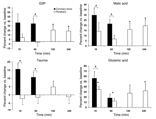
Emerging metabolomic tools have created the opportunity to establish metabolic signatures of myocardial injury. We applied a mass spectrometry-based metabolite profiling platform to 36 patients undergoing alcohol septal ablation treatment for hypertrophic obstructive cardiomyopathy, a human model of planned myocardial infarction (PMI). Serial blood samples were obtained before and at various intervals after PMI, with patients undergoing elective diagnostic coronary angiography and patients with spontaneous myocardial infarction (SMI) serving as negative and positive controls, respectively. We identified changes in circulating levels of metabolites participating in pyrimidine metabolism, the tricarboxylic acid cycle and its upstream contributors, and the pentose phosphate pathway. Alterations in levels of multiple metabolites were detected as early as 10 minutes after PMI in an initial derivation group and were validated in a second, independent group of PMI patients. A PMI-derived metabolic signature consisting of aconitic acid, hypoxanthine, trimethylamine N-oxide, and threonine differentiated patients with SMI from those undergoing diagnostic coronary angiography with high accuracy, and coronary sinus sampling distinguished cardiac-derived from peripheral metabolic changes. Our results identify a role for metabolic profiling in the early detection of myocardial injury and suggest that similar approaches may be used for detection or prediction of other disease states.


Figures

Figure 1
Figure 1. Kinetics of metabolic changes in peripheral human plasma after planned myocardial injury.
Representative metabolites demonstrating transient (≤120 minutes after injury; see Table 2), sustained (≥240 minutes after injury), and late (1,440 minutes after injury, i.e., 1 day) changes in plasma levels compared with baseline are shown. Data represent the entire cohort of 36 patients: boxes denote interquartile range, lines denote median, “+” denotes mean, whiskers denote fifth and ninety-fifth percentiles, and outliers are shown by points.
Figure 2
Figure 2. Kinetic analyses of representative metabolites that are enriched in the coronary sinus after myocardial injury.
Data (mean ± SEM) are restricted to the 13 patients who had simultaneous peripheral blood and coronary sinus sampling performed. Black and white bars show changes in coronary sinus and peripheral levels, respectively. G3P, glycerol-3-phosphate. *P < 0.05 vs. baseline in peripheral blood. #P < 0.05 between coronary sinus and peripheral samples.
Figure 3
Figure 3. Metabolites whose levels significantly changed from baseline at 60, 120, and 240 minutes in PMI patients assessed in an independent SMI cohort.
(A) Black bars denote average of median changes across the 3 time points compared with baseline values in PMI patients. White bars denote relative levels of each metabolite in patients presenting with SMI compared with control patients presenting to the cardiac catheterization suite with nonacute cardiovascular disease. (B) Composite scores for metabolites in A, derived by taking the weighted sum of metabolites that increased in PMI minus the sum of metabolites that decreased in PMI. Boxes denote interquartile range, lines denote median, and whiskers denote tenth and ninetieth percentiles. (C) Receiver-operating-characteristic curve for composite metabolite scores for each patient in the SMI cohort (cases) and the second control cohort (controls). AUC (mean ± SEM) is shown.
Figure 4
Figure 4. Cultured rat cardiomyocytes were subjected to a 3-hour hypoxic challenge as described in Methods.
Cells were incubated with the indicated exogenous metabolites across a range of concentrations. Upon reoxygenation, apoptosis was assessed by FITC-labeled annexin V staining. (A) Percent change in apoptosis relative to the hypoxia control for the lowest concentration of each metabolite screened: xanthine, 14 μM; inosine, 10 μM; glycerol-3-phosphate, 1.7 mM; malonic acid, 6.24 μM; orotic acid, 19 mM; methylhistamine, 1.38 μM; succinic acid, 200 mM; taurine, 2 mM; hypoxanthine, 40 μM. (B) Percent change in apoptosis relative to the hypoxia control at physiologically relevant concentrations of inosine. Representative data (mean ± SEM) from 14 total experiments are shown. *P < 0.05, **P < 0.01 vs. hypoxia control.

References

    1. Nicholson J.K., Wilson I.D. Opinion: understanding ‘global’ systems biology: metabonomics and the continuum of metabolism. Nat. Rev. Drug Discov. 2003;2:668–676. doi: 10.1038/nrd1157. - DOI - PubMed
    1. Raamsdonk L.M., et al. A functional genomics strategy that uses metabolome data to reveal the phenotype of silent mutations. Nat. Biotechnol. 2001;19:45–50. doi: 10.1038/83496. - DOI - PubMed
    1. Allen J., et al. High-throughput classification of yeast mutants for functional genomics using metabolic footprinting. Nat. Biotechnol. 2003;21:692–696. doi: 10.1038/nbt823. - DOI - PubMed
    1. An J., et al. Hepatic expression of malonyl-CoA decarboxylase reverses muscle, liver and whole-animal insulin resistance. Nat. Med. 2004;10:268–274. doi: 10.1038/nm995. - DOI - PubMed
    1. Beecher, C.W.W. 2003. Chap. 17 inThe human metabolome. Kluwer Academic Publishers. Boston, Massachusetts, USA. 311–319.

Publication types

MeSH terms