. 2008 Nov-Dec;14(11-12):697-704.
doi: 10.2119/2008-00072.Gonez. Epub 2008 Aug 20.

Pancreatic expression and mitochondrial localization of the progestin-adipoQ receptor PAQR10

Affiliations

Pancreatic expression and mitochondrial localization of the progestin-adipoQ receptor PAQR10

L Jorge Góñez et al. Mol Med. 2008 Nov-Dec.

Abstract

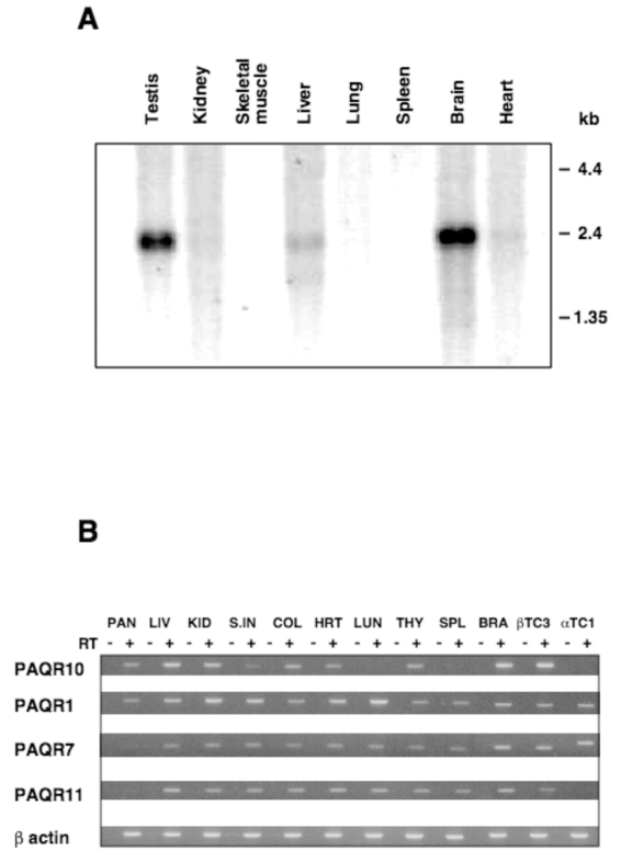
Steroid hormones induce changes in gene expression by binding to intracellular receptors that then translocate to the nucleus. Steroids have also been shown to rapidly modify cell function by binding to surface membrane receptors. We identified a candidate steroid membrane receptor, the progestin and adipoQ receptor (PAQR) 10, a member of the PAQR family, in a screen for genes differentially expressed in mouse pancreatic beta-cells. PAQR10 gene expression was tissue restricted compared with other PAQRs. In the mouse embryonic pancreas, PAQR10 expression mirrored development of the endocrine lineage, with PAQR10 protein expression confined to endocrine islet-duct structures in the late embryo and neonate. In the adult mouse pancreas, PAQR10 was expressed exclusively in islet cells except for its reappearance in ducts of maternal islets during pregnancy. PAQR10 has a predicted molecular mass of 29 kDa, comprises seven transmembrane domains, and, like other PAQRs, is predicted to have an intracellular N-terminus and an extracellular C-terminus. In silico analysis indicated that three members of the PAQR family, PAQRs 9, 10, and 11, have a candidate mitochondrial localization signal (MLS) at the N-terminus. We showed that PAQR10 has a functional N-terminal MLS and that the native protein localizes to mitochondria. PAQR10 is structurally related to some bacterial hemolysins, pore-forming virulence factors that target mitochondria and regulate apoptosis. We propose that PAQR10 may act at the level of the mitochondrion to regulate pancreatic endocrine cell development/survival.


Figures

Figure 1
Figure 1
Tissue expression of PAQR10 mRNA. (A) Northern blotting. A mouse multiple tissue Northern blot (Clontech) was hybridized with a PAQR10-specific cDNA probe as described in Materials and Methods. (B) RT-PCR. Total RNA extracted from different tissues was (+) or was not (−) reverse transcribed and subjected to PCR with specific oligonucleotides for PAQR10, PAQR1, PAQR7, PAQR11, and β-actin (internal control). Amplified products were analyzed on 1.5% agarose gels. PAN, pancreas; LIV, liver; KID, kidney; S.IN, small intestine; COL, colon; HRT, heart; LUN, lung; THY, thymus; SPL, spleen; BRA, brain.
Figure 2
Figure 2
Developmental expression of the PAQR10 gene. Total RNA was extracted from mouse embryos at e10.5 and e12.5 or from dissected pancreas at e13.5, e15.5, e17.5, and newborn stages. RNA was (+) or was not (−) reverse transcribed and subjected to PCR with specific oligonucleotides for PAQR10, Ngn3, PAQR1, PAQR9, PAQR11, and β-actin (internal control). Amplified products were analyzed on 1.5% agarose gels.
Figure 3
Figure 3
Expression of PAQR10 in the developing and adult pancreas. Immunohistochemistry with rat anti-PAQR10 serum was performed on PFA-fixed sections of mouse pancreas, as described in Materials and Methods. Shown are sections of pancreas from e15.5 (10×) (A) and e17.5 (20×) (B) embryos, newborn female mouse (20×) (C), adult female mouse (20×) (D), and e9.5 maternal mouse pancreas at 40× (E) and 100× (F). For controls, maternal mouse pancreas at e9.5 was stained in the absence of primary antibody (G), with rat anti-PAQR10 serum blocked with immunizing peptide (H), or with normal rat serum (I) (all 20×).
Figure 4
Figure 4
Mitochondrial localization of PAQR10. (A) Mitochondrial localization assay. Total (T), cytoplasmic (C), or mitochondria-enriched (M) αTC1 (α) and βTC3 (β) lysates were analyzed for PAQR10 expression by Western blot (left panel). Mitochondrial enrichment was confirmed by Western blot analysis of prohibitin expression in the same C and M lysates (right panel). (B) Confocal microscopy. CHO cells were transfected with plasmid constructs encoding versions of PAQR10 tagged at the C-terminus with myc (PAQR10-myc) or enhanced green fluorescent protein (PAQR10-GFP), at the N-terminus with flag peptide (Flag-PAQR10) or GFP (GFP-PAQR10), or at both ends Flag-PAQR10-myc. Mitochondria were detected by red fluorescence after staining with Mitotracker Red (left panels). PAQR10 or protein tags were detected with the indicated specific antibodies and FITC-labeled secondary antibodies. GFP was visualized under FITC channels (center panels), and the images were merged after confocal microscopy (right panels). These data demonstrate that PAQR10 localizes to mitochondria and that N-terminal tagging of PAQR10 prevents localization and changes its intracellular distribution to perinuclear sites.

References

    1. Watson CS. Signaling themes shared between peptide and steroid hormones at the plasma membrane. Sci STKE. 1999;1999:PE1. - PMC - PubMed
    1. Razandi M, Pedram A, Levin ER. Plasma membrane estrogen receptors signal to antiapoptosis in breast cancer. Mol Endocrinol. 2000;14:1434–47. - PubMed
    1. Simoncini T, et al. Novel non-transcriptional mechanisms for estrogen receptor signaling in the cardiovascular system: interaction of estrogen receptor alpha with phosphatidylinositol 3-OH kinase. Steroids. 2002;67:935–9. - PubMed
    1. Luconi M, et al. Characterization of membrane nongenomic receptors for progesterone in human spermatozoa. Steroids. 2002;67:505–9. - PubMed
    1. Simoncini T, Genazzani AR. Non-genomic actions of sex steroid hormones. Eur J Endocrinol. 2003;148:281–92. - PubMed

Publication types