. 2008 Aug 29;4(8):e1000142.
doi: 10.1371/journal.ppat.1000142.

XIAP regulates cytosol-specific innate immunity to Listeria infection

Affiliations

XIAP regulates cytosol-specific innate immunity to Listeria infection

Laura D Bauler et al. PLoS Pathog. .

Abstract

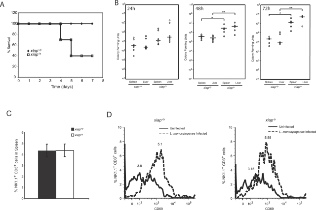
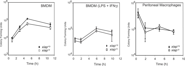
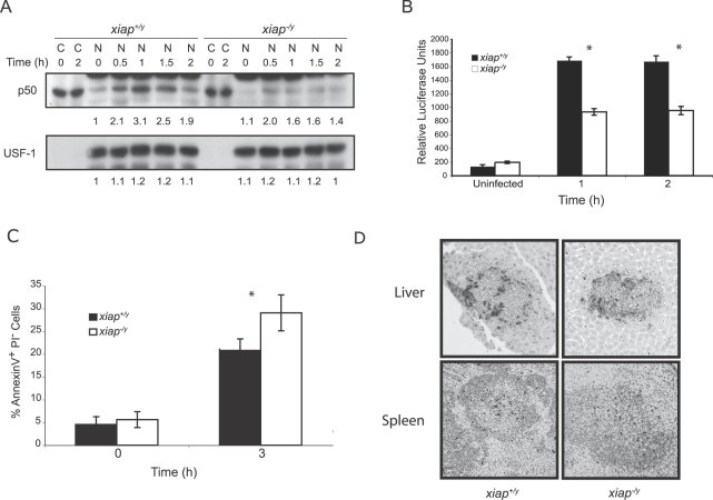
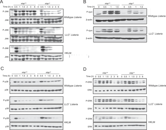
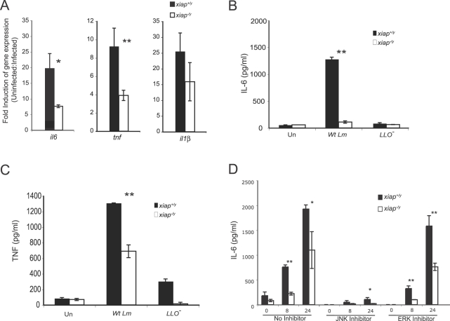
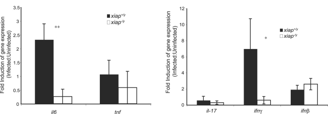
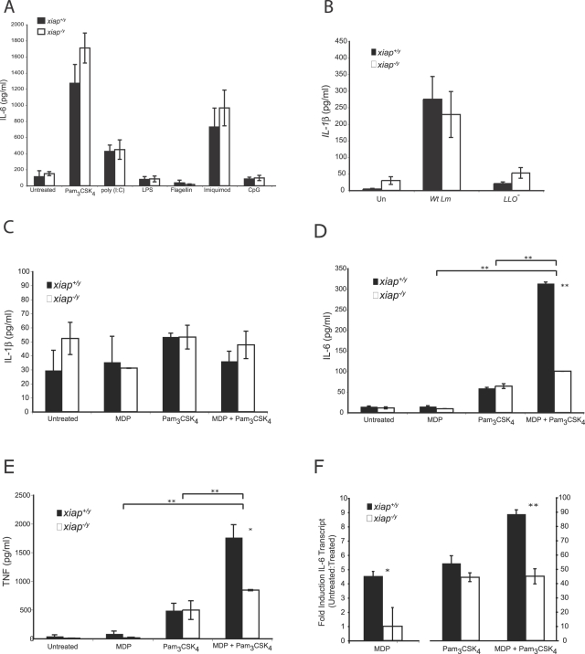
The inhibitor of apoptosis protein (IAP) family has been implicated in immune regulation, but the mechanisms by which IAP proteins contribute to immunity are incompletely understood. We show here that X-linked IAP (XIAP) is required for innate immune control of Listeria monocytogenes infection. Mice deficient in XIAP had a higher bacterial burden 48 h after infection than wild-type littermates, and exhibited substantially decreased survival. XIAP enhanced NF-kappaB activation upon L. monocytogenes infection of activated macrophages, and prolonged phosphorylation of Jun N-terminal kinase (JNK) specifically in response to cytosolic bacteria. Additionally, XIAP promoted maximal production of pro-inflammatory cytokines upon bacterial infection in vitro or in vivo, or in response to combined treatment with NOD2 and TLR2 ligands. Together, our data suggest that XIAP regulates innate immune responses to L. monocytogenes infection by potentiating synergy between Toll-like receptors (TLRs) and Nod-like receptors (NLRs) through activation of JNK- and NF-kappaB-dependent signaling.


Conflict of interest statement

The authors have declared that no competing interests exist.

Figures

Figure 1
Figure 1. XIAP protects against L. monocytogenes infection during the innate immune response.
(A) Survival curve of L. monocytogenes in xiap+/y and xiap−/y mice. Mice were injected with 1×10⁵ L. monocytogenes intraperitoneally, and survival was monitored daily (n = 10 animals per group). (B) CFUs isolated from the liver or spleen of mice infected with 5×10⁵ L. monocytogenes intraperitoneally at 24, 48, and 72 hpi. Each point represents one animal. Mean CFUs is indicated by a horizontal line. *p≤0.05; **p≤0.005. (C) Flow cytometry analysis of NK1.1+CD3+ NKTCs in the spleens of uninfected xiap+/y and xiap−/y animals (error bars represent SD). (D) Activation of NKTCs during L. monocytogenes infection. Splenocytes were harvested from infected animals at 48 hpi, and stained with NK1.1-biotin, CD3-FITC, and CD69-PE fluorescent-coupled antibodies for flow cytometry analysis. Results are representative of three independent experiments (n = 9 animals).
Figure 2
Figure 2. XIAP does not restrict growth of L. monocytogenes in primary macrophages ex vivo.
Intracellular growth of L. monocytogenes in unactivated, activated, and peritoneal macrophages. Unactivated macrophages were infected at an MOI of 1. Activated macrophages were stimulated overnight with 10 ng/ml LPS and 10 ng/ml interferon-γ. Activated and peritoneal macrophages were infected with an MOI of 10 (error bars represent SD).
Figure 3
Figure 3. XIAP enhances NF-κB translocation during L. monocytogenes infection.
(A) Nuclear translocation of p50 in xiap+/y and xiap−/y activated BMDM in response to wild-type L. monocytogenes infection. Cells were activated with 10 ng/ml LPS and 10 ng/ml interferon-γ overnight, and infected at an MOI of 10 for 30 min. Upon lysis, the nuclear fraction (N) was separated by centrifugation from the cytosolic fraction (C). Data are representative of at least 3 independent experiments. (B) DNA binding activity of p50/p65 as measured by ELISA. Nuclear extracts from xiap+/y and xiap−/y activated BMDM that were uninfected or infected with wild-type L. monocytogenes were added to 96-well dishes coated with a canonical NF-κB consensus DNA binding sequence, followed by detection with a p65-specific antibody. Results are representative of at least 3 independent experiments (error bars represent SD). (C) Flow cytometry analysis of apoptosis in activated BMDM infected with L. monocytogenes at 3 hpi. Macrophages were stained at the indicated times post infection with Annexin V-FITC and propidium iodide. Results are representative of at least 3 independent experiments (error bars represent SD of macrophages from 3 mice). (D) TUNEL staining of histological sections of livers and spleens from xiap+/y and xiap−/y mice infected with L. monocytogenes for 48 h (n = 3 animals/genotype). Ten sections per animal were examined.
Figure 4
Figure 4. XIAP prolongs JNK signaling in response to cytosolic L. monocytogenes.
Immunoblot of lysates from xiap+/y and xiap−/y activated BMDM that were uninfected or infected with wild-type, or LLO L. monocytogenes or HKLM. Cells were activated overnight with 10 ng/ml LPS and 10 ng/ml interferon-γ, followed by infection at an MOI of 10 for 30 min. Cells were lysed and subjected to immunoblot analysis using anti-JNK, anti-phospho-JNK, anti-phospho-c-jun, anti-c-jun, anti-phospho-ERK, anti-ERK-1, anti-phospho-p38, and anti-p38 antibodies. Data are representative of at least 3 independent experiments. (A) JNK phosphorylation. (B) c-jun phosphorylation. (C) p38 phosphorylation. (D) ERK phosphorylation. Quantitation of blots can be found in Figure S2.
Figure 5
Figure 5. XIAP regulates proinflammatory cytokine expression upon infection with wild-type L. monocytogenes.
(A) qRT-PCR of genes associated with innate immune activation. BMDM were activated overnight with 10 ng/ml LPS and 10 ng/ml interferon-γ, infected with L. monocytogenes for 30 min, and harvested at 3 hpi for RNA isolation and production of cDNA. Fold induction was calculated using the ΔΔCt method, where uninfected samples were compared to infected samples relative to β-actin levels. (B, C) ELISA of IL-6 (B) and TNF (C) secretion from activated BMDM infected with wild-type or LLO L. monocytogenes. Cells were infected with L. monocytogenes at an MOI of 10 for 30 min. Supernatants were collected at 8 hpi. (D) ELISA of IL-6 secretion from activated BMDM infected with L. monocytogenes and treated with the indicated inhibitors. JNK inhibitor (SP600125) was used at 20 µM, and the ERK inhibitor (U0126) was used at 10 µM. Cells were treated with inhibitors for 1 h, infected at an MOI of 10 for 30 min, and washed with PBS; then, fresh medium with 50 µg/ml gentamicin and the indicated inhibitor was added. Supernatants were collected at 8 and 24 hpi. Error bars represent the SD of macrophages from 3 animals. Results are representative of at least 3 independent experiments. *p≤0.05; **p≤0.005.
Figure 6
Figure 6. In vivo L. monocytogenes infection induces XIAP-dependent pro-inflammatory cytokine expression.
qRT-PCR of genes associated with innate immune activation. Mice were infected with L. monocytogenes, and splenocytes were harvested at 48 hpi for RNA isolation and production of cDNA. Fold induction was calculated using the ΔΔCt method, where uninfected samples were compared to infected samples relative to β2M levels. *p≤0.05; **p≤0.005.
Figure 7
Figure 7. XIAP enables synergistic cytokines responses to TLR and NLR ligands.
(A) IL-6 secretion from xiap+/y and xiap−/y activated BMDM treated with the indicated TLR ligands as measured by ELISA. Macrophages were activated overnight with 10 ng/ml LPS and 10 ng/ml interferon-γ. Cells were left untreated or were treated for 24 h with Pam3CSK4 (2 µg/ml), poly (I:C) (10 µg/ml), LPS (10 ng/ml), flagellin (10 ng/ml), imiquimod (5 µg/ml), or CpG DNA (1 µg/ml). Results are representative of at least 3 independent experiments (error bars represent SD). (B) IL-1β from the supernatants of xiap+/y and xiap−/y activated BMDM left uninfected or infected with wild-type or LLO L. monocytogenes as measured by ELISA. Supernatants were collected at 8 hpi. Results are representative of 3 independent experiments (error bars represent SEM of cells from 6 animals). (C–E) ELISA of IL-1β (C), IL-6 (D), or TNF (E) secretions from xiap+/y and xiap−/y activated BMDM left untreated or treated for 8 h with MDP (10 µg/ml) and/or Pam3CSK4 (0.5 µg/ml). (F) QRTPCR analysis of IL-6 gene expression at 3 h in xiap+/y and xiap−/y activated BMDM treated with MDP (10 µg/ml) and/or Pam3CSK4 (0.5 µg/ml). The data shown are from the same experiment, but are represented on different graphs to show y values more accurately. Data are representative of 3 independent experiments with 3 mice each (error bars represent SD). *p≤0.05; **p≤0.005.

Similar articles

Cited by

References

    1. Salvesen GS, Duckett CS. IAP proteins: blocking the road to death's door. Nat Rev Mol Cell Biol. 2002;3:401–410. - PubMed
    1. Eckelman BP, Salvesen GS, Scott FL. Human inhibitor of apoptosis proteins: why XIAP is the black sheep of the family. EMBO Rep. 2006;7:988–994. - PMC - PubMed
    1. Deveraux QL, Takahashi R, Salvesen GS, Reed JC. X-linked IAP is a direct inhibitor of cell-death proteases. Nature. 1997;388:300–304. - PubMed
    1. Leulier F, Lhocine N, Lemaitre B, Meier P. The Drosophila inhibitor of apoptosis protein DIAP2 functions in innate immunity and is essential to resist gram-negative bacterial infection. Mol Cell Biol. 2006;26:7821–7831. - PMC - PubMed
    1. Kleino A, Valanne S, Ulvila J, Kallio J, Myllymaki H, et al. Inhibitor of apoptosis 2 and TAK1-binding protein are components of the Drosophila Imd pathway. EMBO J. 2005;24:3423–3434. - PMC - PubMed

Publication types

MeSH terms