. 2008 Aug 29;3(8):e3085.
doi: 10.1371/journal.pone.0003085.

Formation of the food vacuole in Plasmodium falciparum: a potential role for the 19 kDa fragment of merozoite surface protein 1 (MSP1(19))

Affiliations

Formation of the food vacuole in Plasmodium falciparum: a potential role for the 19 kDa fragment of merozoite surface protein 1 (MSP1(19))

Anton R Dluzewski et al. PLoS One. .

Abstract

Plasmodium falciparum Merozoite Surface Protein 1 (MSP1) is synthesized during schizogony as a 195-kDa precursor that is processed into four fragments on the parasite surface. Following a second proteolytic cleavage during merozoite invasion of the red blood cell, most of the protein is shed from the surface except for the C-terminal 19-kDa fragment (MSP1(19)), which is still attached to the merozoite via its GPI-anchor. We have examined the fate of MSP1(19) during the parasite's subsequent intracellular development using immunochemical analysis of metabolically labeled MSP1(19), fluorescence imaging, and immuno-electronmicroscopy. Our data show that MSP1(19) remains intact and persists to the end of the intracellular cycle. This protein is the first marker for the biogenesis of the food vacuole; it is rapidly endocytosed into small vacuoles in the ring stage, which coalesce to form the single food vacuole containing hemozoin, and persists into the discarded residual body. The food vacuole is marked by the presence of both MSP1(19) and the chloroquine resistance transporter (CRT) as components of the vacuolar membrane. Newly synthesized MSP1 is excluded from the vacuole. This behavior indicates that MSP1(19) does not simply follow a classical lysosome-like clearance pathway, instead, it may play a significant role in the biogenesis and function of the food vacuole throughout the intra-erythrocytic phase.


Conflict of interest statement

Competing Interests: The authors have declared that no competing interests exist.

Figures

Figure 1
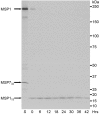
Figure 1. Radiolabeling of MSP119, the final processed fragment of MSP1, showing that it remains attached to the parasite after merozoite invasion of erythrocytes, and that it persists within the parasite throughout its life cycle.
P. falciparum schizonts were radiolabeled with 35S Met/Cys at 45 hours post-invasion for 1.5 hour and a proportion of these parasites were harvested (S, schizonts). The remaining parasites were allowed to undergo invasion, synchronized (2-hour window) and cultured for the next intraerythrocytic cycle. Parasites were harvested every 6 hours from 0 to 42 hours. Rabbit anti-MSP1-specific antibodies were used to immunoprecipitate labeled protein from NP40-extracts of these parasites. The resulting precipitates were analyzed by SDS-PAGE using a 5–12.5% gradient gel under reducing conditions and visualized by fluorography using X-ray film. The polyclonal anti-MSP1 serum recognized the full-length precursor as indicated by the top arrow in the labeled schizont extract, (S), and the 19-kDa fragment in newly formed ring stages (0 hours), indicated by the bottom arrow. The processed fragment of MSP7, MSP733 is also detected as part of the MSP1 complex in schizonts (middle arrow). The remaining lanes represent parasites harvested every 6 hours from 6 to 42 hours post-invasion. The intensity and the mobility of the MSP119 band remained constant until 42 hours when there was some decrease in intensity; this reduction in intensity could be due to rupture of some mature forms releasing MSP119 into the culture medium. The mobility of protein size markers is indicated at the right of the panel according to their size (kDa).
Figure 2
Figure 2. Immunofluorescence microscopy showing the localization of MSP119 and CRT in parasites that had invaded in the presence of mAb 1E1; showing that MSP119 moves to the food vacuole.
Schizonts were allowed to release merozoites that invaded fresh erythrocytes in the presence of mAb 1E1, then ring-stage parasites were cultured and samples harvested every 3 hours over the asexual cycle. The mAb 1E1 antibody associates with MSP119 on the surface of the merozoite, and is detected within the ring-stage parasite; it was used to follow MSP119 in smears counterstained with rabbit anti-MSP119 (panel A) and rabbit anti-CRT (panel B), which is a marker of the food vacuole. Each row of four panels show an identical field from 1% formaldehyde-fixed thin smears of P. falciparum ring-stage parasites; columns A1 and B1 show mAb 1E1 associated with the parasite detected with Alexa Fluor 488 conjugated anti-mouse IgG (green); columns A2 rabbit anti-MSP119 and B2 rabbit anti-CRT, detected with Alexa Fluor 594 conjugated anti-rabbit IgG (red); columns A3 and B3 are composites of columns 1 and 2 with DAPI staining of the nucleus (blue) and any green and red fluorescence overlap is displayed in yellow. In columns A4 and B4, the parasitised erythrocyte is visualized by light microscopy, showing the location of the parasite within the infected erythrocyte. Only five time points are shown: 0, 6, 9, 12 and 18 hours post-invasion. The two anti-MSP119 antibodies, in panels A1 and A2, are seen to co-localize in panel A3, showing that 1E1 is still associated with MSP119 in the young parasite. CRT is clearly detectable from 6 to 9 hours onwards post-invasion (panel B2) and appears to be closely associated with MSP119 (panel B3). Pigment granules are clearly visible by light microscopy from about 18 hours (panels A4 and B4). Similar results were obtained in a separate experiment in which mAb 1E1 was used to detect MSP119 on formaldehyde fixed parasites at the same time points (data not shown) and at 24 hours (panel C). Panels C1–C4 show 1E1, (C1, green), rabbit anti-CRT (C2, red), composites of these antibodies with DAPI staining merged (C3) and light visualization (C4), as described above. CRT was clearly detectable (C2) and largely co-localized with MSP119 (C3). Both MSP119 and CRT were associated with the pigment detected by light microscopy in panel C4 and no longer around the surface of the parasite.
Figure 3
Figure 3. IFA location of MSP119, CRT and new full length MSP1 during schizogony.
Mature parasites at different stages of nuclear division were compared. Each row in panels A and B shows an identical field from 1% formaldehyde-fixed thin smears of late-stages parasites at the 1-, 2-, 4-, and 8-nuclei stage. Panels A1 and B1 show anti-MSP119(rabbit) and rabbit anti-CRT(rabbit) antibody labeling respectively (the secondary antibody was Alexa Fluor 488-conjugated anti-rabbit IgG antibody (green). Panels A2 and B2 show the same fields merged with the corresponding DAPI stained nucleus images (blue). Panels A3 and B3 show the same fields visualized by light microscopy - note the pigment is clearly visible. Rabbit-anti-MSP119 antibody only detects MSP119 associated with the food vacuole until the two-nuclei stage. Once new full length MSP1 is synthesized and transported to the parasite's plasma membrane the presence of MSP119 from the previous cycle is now obscured. In contrast, rabbit-anti-CRT antiobody is clearly detectable throughout parasite maturation and is associated with the food vacuole. Panel C, rows (a) and (b), shows that MSP1 and CRT are associated with the residual body that is released upon schizont rupture. Thin smears made at the time of schizont rupture were probed consecutively with mAb 1E1 and rabbit anti-CRT antibodies. These antibodies were detected using Alexa Fluor 488-conjugated anti-mouse IgG, and Alexa Fluor 594-conjugated anti-rabbit IgG, respectively (columns 1 and 2). Column 4 shows the bright field microscopy images of the same field and clearly indicate the presence of malarial pigment. Column 5 shows the absence of nuclear material in the residual bodies, as confirmed by the lack of DAPI staining. Column 3 shows the merged images from columns 1, 2 and 5 - both CRT and MSP119, or MSP1, which cannot be distinguished by the antibody, are associated with the released pigment in the residual body.
Figure 4
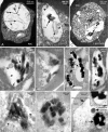
Figure 4. Ultrastructural morphology of endocytic vacuoles in ring stages.
Panels A to G show sections through established ring stages prepared for EM morphology, A and C show the two types of vacuole present in the cytoplasm, one not containing hemoglobin (clear vacuoles) and the other with hemoglobin (small food vacuoles). An obliquely sectioned cytostome and a group of small dense food vacuoles are visible in B. In C a double membrane dense food vacuole is also shown, which is typical of early endocytosis before the inner membrane breaks down (see text). In panels D–F three sections from a serial section sequence are shown, depicting stages in hemozoin formation in a mid-stage ring. In panel D a vacuole in the early stages of hemoglobin degradation is visible (arrow, bottom left) as well as smaller vacuoles containing dense masses indicating hemozoin formation, In panel F a larger vacuole containing a number of small hemozoin crystals is shown (enlarged in panel G). Abbreviations: RER–rough endoplasmic reticulum; RBC–red blood cell.
Figure 5
Figure 5. The distribution of MSP119 in ring-stage parasites detected by IEM.
Panels D and E show MSP119-specific labeling of the surface of early rings, and in panel E, a clear endocytic vacuole is also labeled. Panels F–H show that MSP119 has been endocytosed into the small dense food vacuoles, and is located mainly at the inner vacuolar membrane surface; in Panel G the food vacuoles have a tubular appearance. In Panel I, a late ring is labeled, showing the incorporation of MSP119 into larger vacuoles containing small hemozoin crystals (Hz), with the label again associated with the vacuole membrane. Abbreviations: Fvtu- tubular food vacuoles; Hz- hemozoin.
Figure 6
Figure 6. The distribution of MSP1 within late trophozoites and schizonts.
Panels A–C show the appearance of the single food vacuole, prepared for EM morphology. Panel A shows a late trophozoite containing a typically dilated food vacuole, containing widely spaced hemozoin and several profiles of internal membranous structures. In panel B an early schizont stage vacuole contains some hemozoin crystals and internal membrane profiles, while the vacuole wall shows signs of inward folding (arrowheads). C shows another schizont where a lipid body has formed adjacent to the food vacuole. Panels D–G show specific immunogold labeling of early to mid-stage schizonts labeled with MSP119-specific antibodies, in all cases detecting the protein at the vacuole wall. In panel D a limited area of labeling is present along one side of the food vacuole (small arrows), and in E a similar distribution is seen on a more folded vacuole wall. Panels F and G show labeling along obliquely sectioned folds of the wall (arrows), where the label also lies close to hemozoin crystals. Panels H and I show food vacuoles of late-stage schizonts containing closely packed large hemozoin crystals and are almost completely surrounded by MSP119 labeled vacuole wall membrane. In Panel J (and the inset of a portion at higher magnification) an early-/mid-stage schizont has been immunostained with antibody reacting with N-terminal regions of MSP1 but not MSP119. This antibody labels the newly synthesized (full-length) MSP1 on the parasite's plasma membrane but fails to label the food vacuole, indicating that the MSP119-specific labeling, seen in panels D–I, is specific for MSP119 carried in on the parasite surface at invasion rather than new MSP1 recently synthesized by the schizont. Abbreviations: FV–food vacuole; Hz–hemozoin; Int mem–internal membranous structures; RBC–red blood cell.
Figure 7
Figure 7. A diagram summarising the general fate of MSP1 and the C-terminal fragment MSP119 through the asexual cycle.
In panel A, MSP1 on the surface of the invading merozoite undergoes cleavage to release the N-terminal portion (orange) and leave the C-terminal MSP119 (green) attached to the surface of the parasite: firstly as a newly internalized merozoite; and later as it transforms into a ring stage. MSP119 is then endocytosed from the ring surface into small food vacuoles as the parasite begins to feed on and metabolise hemoglobin. In B, all the MSP119 has been endocytosed and is associated with the membrane of the now single food vacuole, where it remains through the mid-schizont stage (C) and is then incorporated into the residual body at the end of schizogony (D).
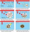
Figure 8
Figure 8. A series of diagrams summarising in greater detail the main conclusions relating to post-invasion MSP119 endocytosis and its progression through the asexual cycle.
In each panel a portion of the parasite is shown in blue, with the adjacent RBC in red, and with the parasitophorous vacuole space between the two. The fragments of MSP1 are colored as in Figure 7.

Similar articles

Cited by

References

    1. Blackman MJ, Heidrich HG, Donachie S, McBride JS, Holder AA. A single fragment of a malaria merozoite surface protein remains on the parasite during red cell invasion and is the target of invasion-inhibiting antibodies. J Exp Med. 1990;172:379–382. - PMC - PubMed
    1. Hall R, McBride J, Morgan G, Tait A, Zolg JW, et al. Antigens of the erythrocytes stages of the human malaria parasite Plasmodium falciparum detected by monoclonal antibodies. Mol Biochem Parasitol. 1983;7:247–265. - PubMed
    1. Gilson PR, O'Donnell RA, Nebl T, Sanders PR, Wickham ME, et al. MSP1(19) miniproteins can serve as targets for invasion inhibitory antibodies in Plasmodium falciparum provided they contain the correct domains for cell surface trafficking. Mol Microbiol. 2008;68:124–138. - PubMed
    1. Oka M, Aikawa M, Freeman RR, Holder AA, Fine E. Ultrastructural localization of protective antigens of Plasmodium yoelii merozoites by the use of monoclonal antibodies and ultrathin cryomicrotomy. Am J Trop Med Hyg. 1984;33:342–346. - PubMed
    1. Heidrich HG, Matzner M, Miettinen-Baumann A, Strych W. Immunoelectron microscopy shows that the 80,000-dalton antigen of Plasmodium falciparum merozoites is localized in the surface coat. Z Parasitenkd. 1986;72:681–683. - PubMed

Publication types

MeSH terms