. 2008 Sep;79(9):1679-88.
doi: 10.1902/jop.2008.080024.

Modeling of trabecular bone and lamina dura following selective alveolar decortication in rats

Affiliations

Modeling of trabecular bone and lamina dura following selective alveolar decortication in rats

Jean-David Sebaoun et al. J Periodontol. 2008 Sep.

Abstract

Background: Modifying the balance between resorption and apposition through selectively injuring the cortical plate of the alveolus has been an approach to speed tooth movement and is referred to as periodontally accelerated osteogenic orthodontics. The aim of this study was to investigate the alveolar response to corticotomy as a function of time and proximity to the surgical injury in a rat model.

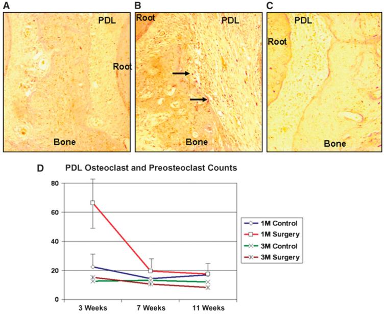
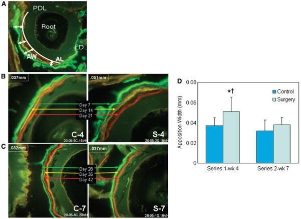
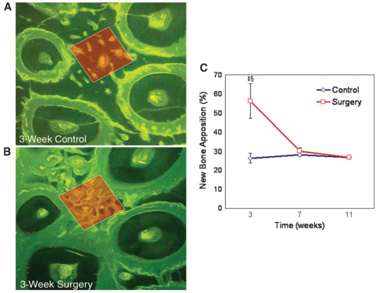
Methods: Maxillary buccal and lingual cortical plates were injured in 36 healthy adult rats adjacent to the upper left first molars. Twenty-four animals were euthanized at 3, 7, or 11 weeks. In one group, the maxillae were removed and stripped of soft tissues, and histomorphometric analysis was performed to study alveolar spongiosa and periodontal ligament (PDL) modeling dynamics. Catabolic activity was analyzed with tartrate-resistant acid phosphatase-positive osteoclasts and preosteoclasts. Anabolic actions were measured using a fluorescent vital bone stain series followed by sacrifice at 30 and 51 days. To further analyze the new bone formation, a separate group of animals were fed with calcein fluorescent stain and processed for non-decalcified fluorescent stain histology.

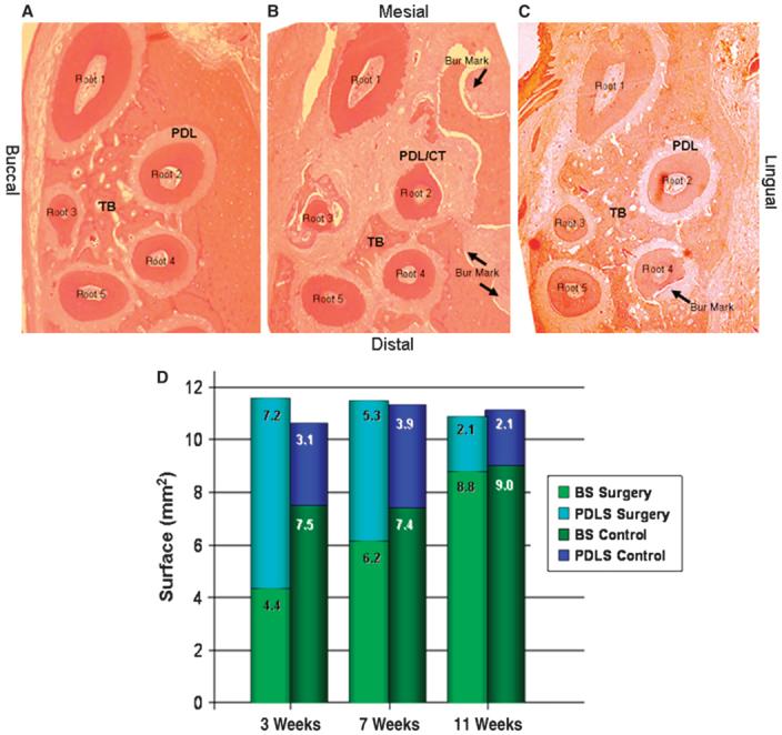
Results: At 3 weeks, the surgery group had significantly (P <0.05) less calcified spongiosa bone surface, greater periodontal ligament surface, higher osteoclast number, and greater lamina dura apposition width. The catabolic activity (osteoclast count) and anabolic activity (apposition rate) were three-fold greater, calcified spongiosa decreased by two-fold, and PDL surface increased by two-fold. Surgical injury to the alveolus that induced a significant increase in tissue turnover by week 3 dissipated to a steady state by postoperative week 11. The impact of the injury was localized to the area immediately adjacent to the decortication injury.

Conclusion: Selective alveolar decortication induced increased turnover of alveolar spongiosa, and the activity was localized; dramatic escalation of demineralization-remineralization dynamics is the likely biologic mechanism underlying rapid tooth movement following selective alveolar decortication.


Figures

Figure 1
Figure 1
Surgical procedure and analysis. A) The corticotomy procedure. Red and blue circles indicate surgical bur cuts on buccal and lingual surfaces, respectfully. B) Histomorphometric analysis was performed for bone and PDL modeling dynamics and osteoclast count (hematoxylin and eosin; original magnification: ×2.5). CP = cortical plate; TB = trabecular bone.
Figure 2
Figure 2
Histopathologic analysis of tissue response. The contralateral control sites (A) had significantly more trabecular bone surface compared to the surgery group at 3 weeks (B). There was a significantly greater PDL surface in the surgery group at the third week compared to the control group. C) Eleven weeks after surgery, the tissue architecture was completely restored and returned to baseline. (Hematoxylin and eosin; original magnification: A through C, ×2.5.) CT = connective tissue; TB = trabecular bone. Arrows indicate the bur marks; root 1 through root 5 denote the roots of the first maxillary molar. D) The surface area of the trabecular bone (BS) and the PDL (PDLS; the extended PDL area into the connective tissue that replaced the resorbing bone) were inversely correlated; the smallest amount of trabecular bone surface was observed at the 3-week analysis. By the end of 11 weeks, the proportional distribution of the BS and the PDLS at the surgery site was similar to the contralateral side.
Figure 3
Figure 3
Osteoclastic activity and bone resorption. The number of TRAP-positive osteoclasts and preosteoclasts was significantly lower in the control group (A) compared to the surgery group (B) by the end of 3 weeks. C) Eleven weeks after the surgery, the TRAP-positive cell counts and their distribution at the surgery site were similar to baseline and the contralateral control sides. Arrows indicate TRAP-positive osteoclasts. (TRAP; original magnification: A through C, ×20.) D) The first-molar (lM) PDL of the surgery group at 3 weeks had significantly greater osteoclastic activity than the 3-week control and the other lM groups, as well as the third-molar (3M) areas at 3, 7, and 11 weeks. M = molar.
Figure 4
Figure 4
Lamina dura apposition width. A) Following injection of calcein (green), tetracycline (yellow), and alizarin red (red), apposition length (AL; curved line) and apposition width (AW; arrows) of the lamina dura (LD) surrounding the root were measured. B) After series 1 injections at 7, 14, and 21 days and sacrifice at 30 days, lamina dura width around the first molar was significantly greater on the decortication side (S-4) compared to the control (C-4). C) After series 2 injections at 28, 35, and 42 days and sacrifice at 51 days, there were no significant differences between decortication (S-7) and control sides (C-7), whereas the apposition width at 4 weeks was significantly higher than the surgery group at 7 weeks. (Original magnification: A through C, ×20.) D) Apposition width of lamina dura as a function of time in surgery and control groups (mean ± SEM). *P <0.001 compared to control at week 4; P <0.001 compared to surgery at week 7. wk = week.
Figure 5
Figure 5
New trabecular bone formation. The percentage of new bone apposition in the first-molar area was significantly lower in the control sites (A) compared to the postdecortication group at 3 weeks (B) (calcein ad libitum; original magnification: ×10). C) Anabolic modeling of alveolar trabecular bone adjacent to the decortication site increased by ∼1.5 times at 3 weeks; this change was diminished by 7 weeks to the level of the control side and was stabilized by 11 weeks (mean ± SEM). P <0.01 compared to control at week 3; P <0.01 compared to surgery at weeks 7 and 11.

References

    1. Roberts WE, Huja S, Roberts JA. Bone modeling: Biomechanics, molecular mechanisms, and clinical perspectives. Semin Orthod. 2004;10:123–161.
    1. Verna C, Dalstra M, Melsen B. The rate and the type of orthodontic tooth movement is influenced by bone turnover in a rat model. Eur J Orthod. 2000;22:343–352. - PubMed
    1. Midgett RJ, Shaye R, Fruge JF., Jr. The effect of altered bone metabolism on orthodontic tooth movement. Am J Orthod. 1981;80:256–262. - PubMed
    1. Goldie RS, King GJ. Root resorption and tooth movement in orthodontically treated, calcium-deficient, and lactating rats. Am J Orthod. 1984;85:424–430. - PubMed
    1. Engström C, Granström G, Thilander B. Effect of orthodontic force on periodontal tissue metabolism. A histologic and biochemical study in normal and hypocalcemic young rats. Am J Orthod Dentofacial Orthop. 1988;93:486–495. - PubMed

Publication types

MeSH terms