

Phagocytic clearance of electric field induced 'apoptosis-mimetic' cells
- PMID: 18771656
- PMCID: PMC2716758
- DOI: 10.1016/j.bbrc.2008.08.060
Phagocytic clearance of electric field induced 'apoptosis-mimetic' cells
Abstract
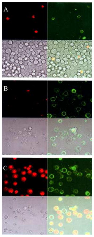
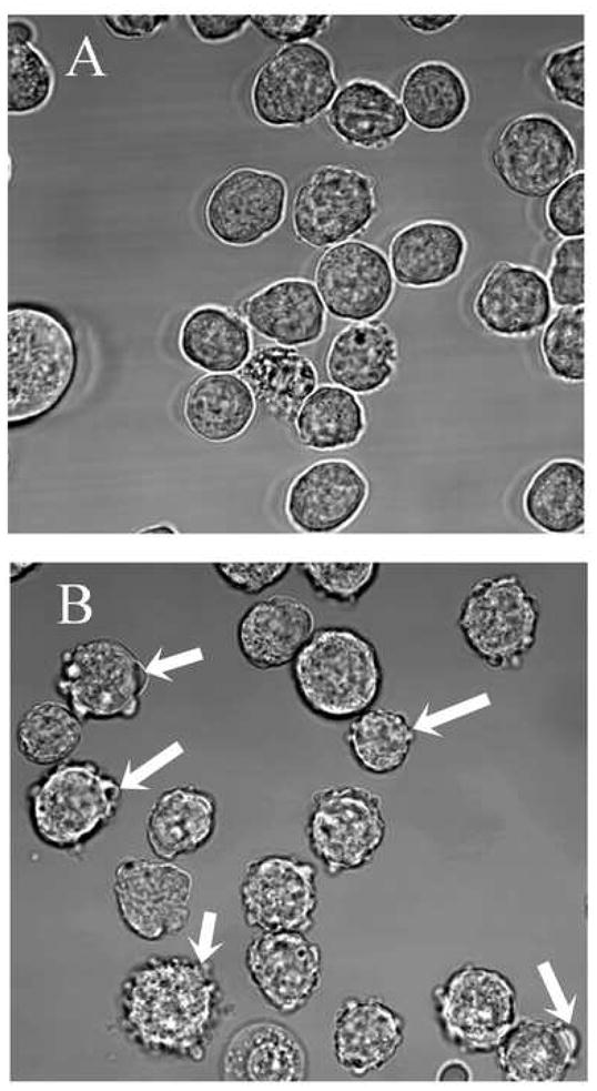
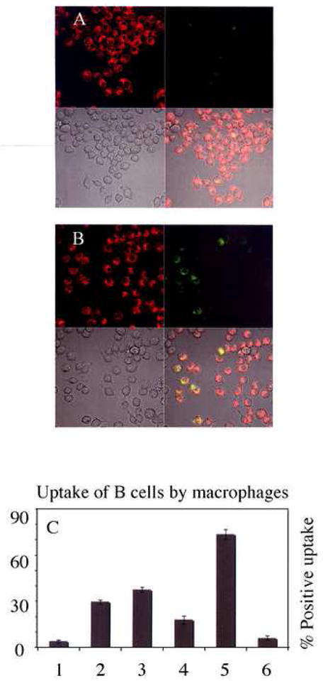
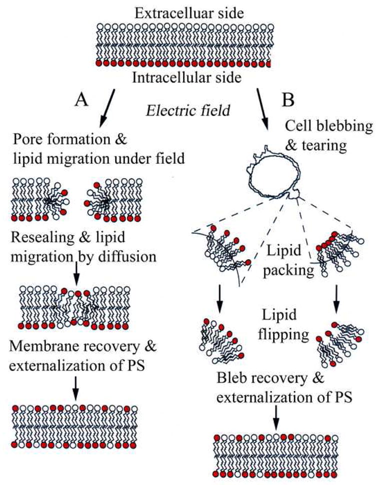
Cells undergoing apoptosis lose lipid asymmetry that is often manifested by the exposure of phosphatidylserine (PS) to the outer surface of the cell membrane. Macrophages and other cell types recognize externalized PS to signal phagocytosis, thereby eliciting a non-inflammatory response. PS exposure is obligatory in the recognition and clearance of apoptotic cells. Here, we find that externally applied moderate electric field induces PS externalization in a mouse B-cell (FOX-NY) membrane without procaspase-3 activation, a major characteristic of apoptotic cells. The field-induced PS inversion is caused as a result of electroporation and/or a process involving membrane reorganizations and recovery that ensues following field exposure. Using a mouse macrophage cell line (J7444A.1) from the same strain, we show phagocytic clearance of PS expressing B-cells and demonstrate that this is in part due to the apoptosis mimicry of the field exposed cells.
Figures
Similar articles
-
Macrophage recognition of externalized phosphatidylserine and phagocytosis of apoptotic Jurkat cells--existence of a threshold.Arch Biochem Biophys. 2003 May 1;413(1):41-52. doi: 10.1016/s0003-9861(03)00083-3. Arch Biochem Biophys. 2003. PMID: 12706340
-
A role for oxidative stress in apoptosis: oxidation and externalization of phosphatidylserine is required for macrophage clearance of cells undergoing Fas-mediated apoptosis.J Immunol. 2002 Jul 1;169(1):487-99. doi: 10.4049/jimmunol.169.1.487. J Immunol. 2002. PMID: 12077280
-
Essential role of phosphatidylserine externalization in apoptosing cell phagocytosis by macrophages.Biochem Biophys Res Commun. 1998 May 19;246(2):549-55. doi: 10.1006/bbrc.1998.8663. Biochem Biophys Res Commun. 1998. PMID: 9610400
-
The central role of phosphatidylserine in the phagocytosis of apoptotic thymocytes.Ann N Y Acad Sci. 2000;926:217-25. doi: 10.1111/j.1749-6632.2000.tb05614.x. Ann N Y Acad Sci. 2000. PMID: 11193037 Review.
-
Phosphatidylserine, a death knell.Cell Death Differ. 2001 Jun;8(6):551-63. doi: 10.1038/sj.cdd.4400817. Cell Death Differ. 2001. PMID: 11536005 Review.
Cited by
-
Electrical Stimulation for Immunomodulation.ACS Omega. 2023 Dec 20;9(1):52-66. doi: 10.1021/acsomega.3c06696. eCollection 2024 Jan 9. ACS Omega. 2023. PMID: 38222551 Free PMC article. Review.
-
Whole blood cells loaded with messenger RNA as an anti-tumor vaccine.Adv Healthc Mater. 2014 Jun;3(6):837-42. doi: 10.1002/adhm.201300512. Epub 2013 Dec 16. Adv Healthc Mater. 2014. PMID: 24339387 Free PMC article.
-
Cellular regulation of extension and retraction of pseudopod-like blebs produced by nanosecond pulsed electric field (nsPEF).Cell Biochem Biophys. 2014 Jul;69(3):555-66. doi: 10.1007/s12013-014-9831-9. Cell Biochem Biophys. 2014. PMID: 24488232 Free PMC article.
-
Starting a Fire Without Flame: The Induction of Cell Death and Inflammation in Electroporation-Based Tumor Ablation Strategies.Front Oncol. 2020 Jul 28;10:1235. doi: 10.3389/fonc.2020.01235. eCollection 2020. Front Oncol. 2020. PMID: 32850371 Free PMC article. Review.
-
Distinct roles but cooperative effect of TLR3/9 agonists and PD-1 blockade in converting the immunotolerant microenvironment of irreversible electroporation-ablated tumors.Cell Mol Immunol. 2021 Dec;18(12):2632-2647. doi: 10.1038/s41423-021-00796-4. Epub 2021 Nov 15. Cell Mol Immunol. 2021. PMID: 34782757 Free PMC article.
References
-
- Bevers EM, Comfurius P, Zwaal RF. Changes in membrane phospholipid distribution during platelet activation. Biochim Biophys Acta. 1983;736:57–66. - PubMed
-
- Wali RK, Jaffe S, Kumar D, Kalra VK. Alternations in organization of phospholipids in erythrocytes as a factor in adherence to endothelial cells in diabetes mellitus. Diabetes. 1988;37:104–111. - PubMed
-
- Erwig LP, Henson PM. Clearance of apoptotic cells by phagocytes. Cell Death Differ. 2008;15:243–250. - PubMed
-
- Fadok VA, Voelker DR, Campbell PA, Cohen JJ, Bratton DL, Henson PM. Exposure of phosphatidylserine on the surface apoptotic lymphocytes triggers specific recognition and removal by macrophages. J Immunol. 1992;148:2207–2216. - PubMed
-
- Fadok VA, de Cathelineau A, Daleke DL, Henson PM, Bratton DL. Loss of phospholipid asymmetry and surface exposure of phosphatidylserine is required for phagocytosis of apoptotic cells by macrophages and fibroblasts. J Biol Chem. 2001;276:1071–1077. - PubMed
Publication types
MeSH terms
Substances
Grants and funding
LinkOut - more resources
Full Text Sources
Research Materials
