

Androgen and estrogen (alpha) receptor localization on periaqueductal gray neurons projecting to the rostral ventromedial medulla in the male and female rat
- PMID: 18771723
- PMCID: PMC2626772
- DOI: 10.1016/j.jchemneu.2008.08.001
Androgen and estrogen (alpha) receptor localization on periaqueductal gray neurons projecting to the rostral ventromedial medulla in the male and female rat
Abstract
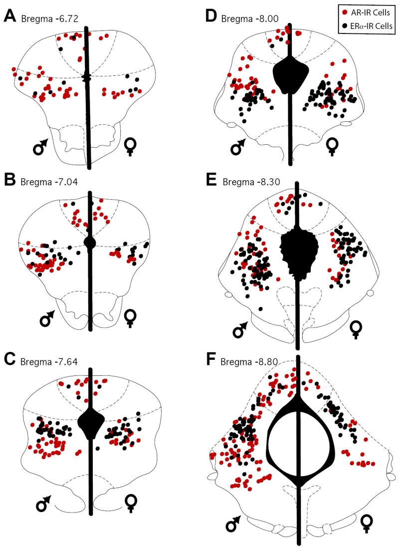
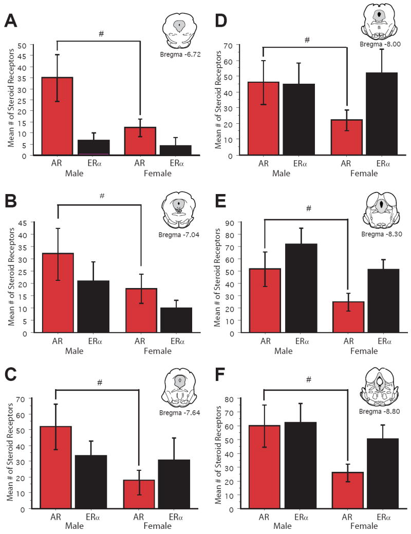
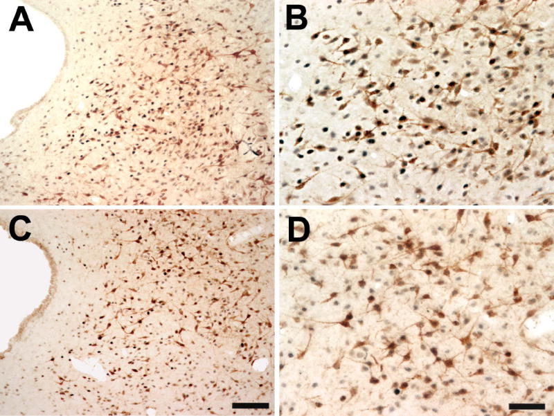
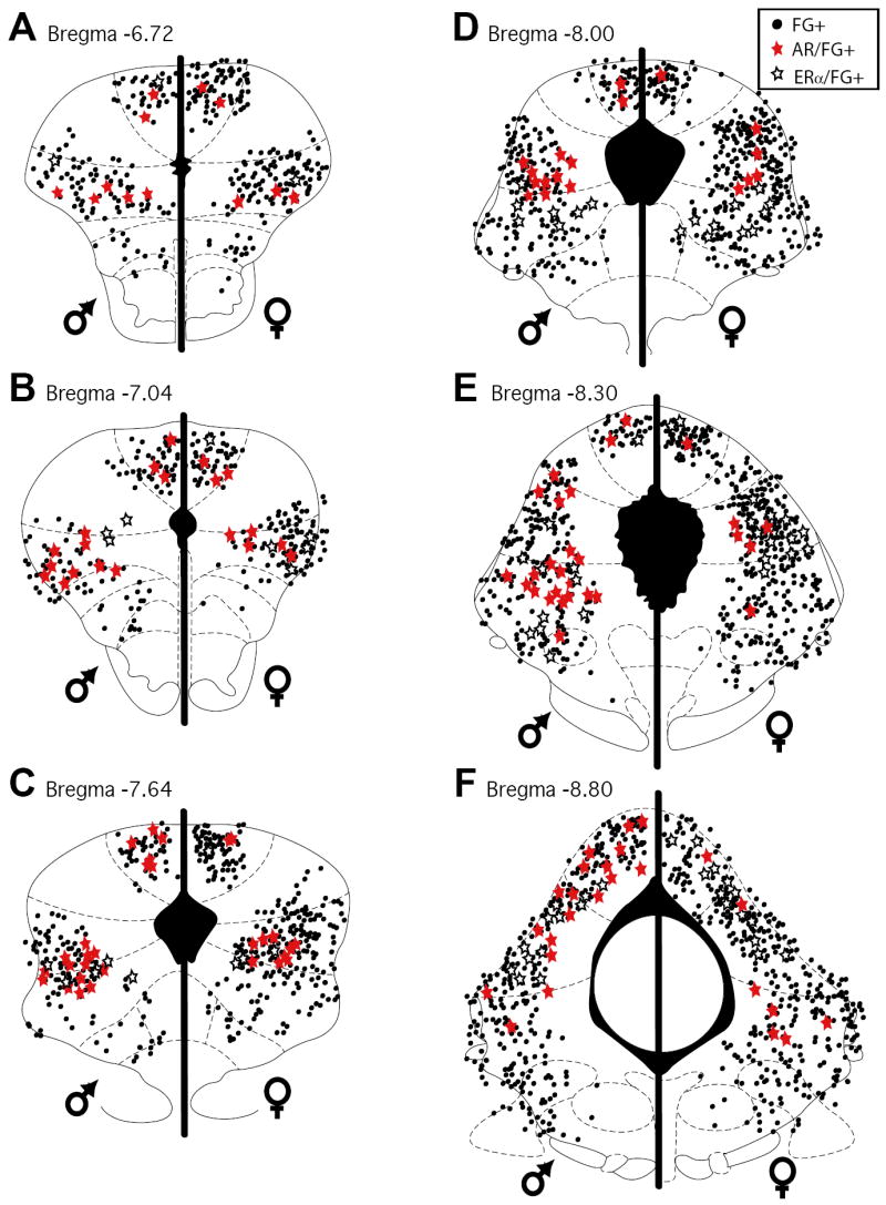
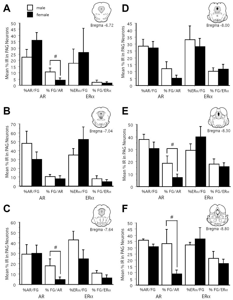
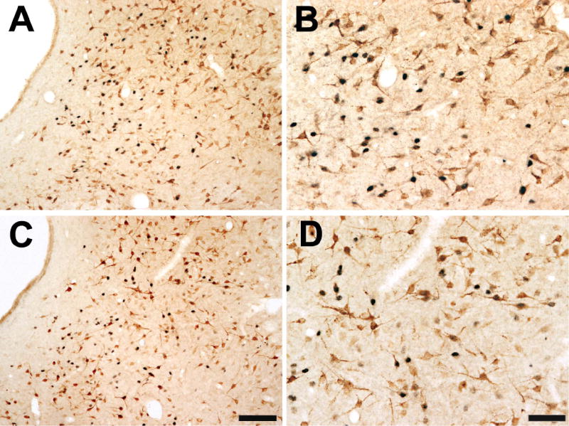
The periaqueductal gray (PAG) is involved in many gonadal steroid-sensitive behaviors, including responsiveness to pain. The PAG projects to the rostral ventromedial medulla (RVM), comprising the primary circuit driving pain inhibition. Morphine administered systemically or directly into the PAG produces greater analgesia in male compared to female rats, while manipulation of gonadal hormones alters morphine potency in both sexes. It is unknown if these alterations are due to steroidal actions on PAG neurons projecting to the RVM. The expression of androgen (AR) and estrogen (ERalpha) receptors in the PAG of female rats and within this descending inhibitory pathway in both sexes is unknown. The present study used immunohistochemical techniques (1) to map the distribution of AR and ERalpha across the rostrocaudal axis of the PAG; and (2) to determine whether AR and/or ERalpha were colocalized on PAG neurons projecting to the RVM in male and female rats. AR and ERalpha immunoreactive neurons (AR-IR, ERalpha-IR) were densely distributed within the caudal PAG of male rats, with the majority localized in the lateral/ventrolateral PAG. Females had significantly fewer AR-IR neurons, while the quantity of ERalpha was comparable between the sexes. In both sexes, approximately 25-50% of AR-IR neurons and 20-50% of ERalpha-IR neurons were retrogradely labeled. This study provides direct evidence of the expression of steroid receptors in the PAG and the descending pathway driving pain inhibition in both male and female rats and may provide a mechanism whereby gonadal steroids modulate pain and morphine potency.
Figures
Similar articles
-
Mu- and delta-opioid receptor mRNAs are expressed in periaqueductal gray neurons projecting to the rostral ventromedial medulla.Neuroscience. 2002;109(3):619-34. doi: 10.1016/s0306-4522(01)00328-1. Neuroscience. 2002. PMID: 11823071
-
Morphine preferentially activates the periaqueductal gray-rostral ventromedial medullary pathway in the male rat: a potential mechanism for sex differences in antinociception.Neuroscience. 2007 Jun 29;147(2):456-68. doi: 10.1016/j.neuroscience.2007.03.053. Epub 2007 May 31. Neuroscience. 2007. PMID: 17540508 Free PMC article.
-
Androgen and estrogen (alpha) receptor distribution in the periaqueductal gray of the male Rat.Horm Behav. 1999 Oct;36(2):98-108. doi: 10.1006/hbeh.1999.1528. Horm Behav. 1999. PMID: 10506534
-
The role of the periaqueductal gray in the modulation of pain in males and females: are the anatomy and physiology really that different?Neural Plast. 2009;2009:462879. doi: 10.1155/2009/462879. Epub 2009 Jan 28. Neural Plast. 2009. PMID: 19197373 Free PMC article. Review.
-
Neuronal and glial factors contributing to sex differences in opioid modulation of pain.Neuropsychopharmacology. 2019 Jan;44(1):155-165. doi: 10.1038/s41386-018-0127-4. Epub 2018 Jun 23. Neuropsychopharmacology. 2019. PMID: 29973654 Free PMC article. Review.
Cited by
-
Function of gastrin-releasing peptide receptors in ocular itch transmission in the mouse trigeminal sensory system.Front Mol Neurosci. 2023 Nov 30;16:1280024. doi: 10.3389/fnmol.2023.1280024. eCollection 2023. Front Mol Neurosci. 2023. PMID: 38098939 Free PMC article.
-
Interactive Mechanisms of Supraspinal Sites of Opioid Analgesic Action: A Festschrift to Dr. Gavril W. Pasternak.Cell Mol Neurobiol. 2021 Jul;41(5):863-897. doi: 10.1007/s10571-020-00961-9. Epub 2020 Sep 24. Cell Mol Neurobiol. 2021. PMID: 32970288 Free PMC article. Review.
-
Testosterone protects against the development of widespread muscle pain in mice.Pain. 2020 Dec;161(12):2898-2908. doi: 10.1097/j.pain.0000000000001985. Pain. 2020. PMID: 32658149 Free PMC article.
-
Testosterone modulation of ethanol effects on the µ-opioid receptor kinetics in castrated rats.Biomed Rep. 2019 Sep;11(3):103-109. doi: 10.3892/br.2019.1230. Epub 2019 Jul 18. Biomed Rep. 2019. PMID: 31423304 Free PMC article.
-
Proposing a neural framework for the evolution of elaborate courtship displays.Elife. 2022 May 31;11:e74860. doi: 10.7554/eLife.74860. Elife. 2022. PMID: 35639093 Free PMC article.
References
-
- Albert DJ, Jonik RH, Walsh ML. Hormone-dependent aggression in the female rat: testosterone plus estradiol implants prevent the decline in aggression following ovariectomy. Physiol Behav. 1991;49:673–677. - PubMed
-
- Albert DJ, Jonik RH, Watson NV, Gorzalka BB, Walsh ML. Hormone-dependent aggression in male rats is proportional to serum testosterone concentration but sexual behavior is not. Physiol Behav. 1990;48:409–416. - PubMed
-
- Aloisi AM, Ceccarelli I, Fiorenzani P, De Padova AM, Massafra C. Testosterone affects formalin-induced responses differently in male and female rats. Neurosci Lett. 2004;361:262–264. - PubMed
-
- Alper RH, Schmitz TM. Estrogen increases the bradycardia elicited by central administration of the serotonin1A agonist 8-OH-DPAT in conscious rats. Brain Res. 1996;716:224–228. - PubMed
-
- Bandler R, Carrive P. Integrated defence reaction elicited by excitatory amino acid microinjection in the midbrain periaqueductal grey region of the unrestrained cat. Brain Res. 1988;439:95–106. - PubMed
Publication types
MeSH terms
Substances
Grants and funding
LinkOut - more resources
Full Text Sources
Research Materials
