

Protein adsorption and cell adhesion on nanoscale bioactive coatings formed from poly(ethylene glycol) and albumin microgels
- PMID: 18771802
- PMCID: PMC2633218
- DOI: 10.1016/j.biomaterials.2008.08.003
Protein adsorption and cell adhesion on nanoscale bioactive coatings formed from poly(ethylene glycol) and albumin microgels
Abstract
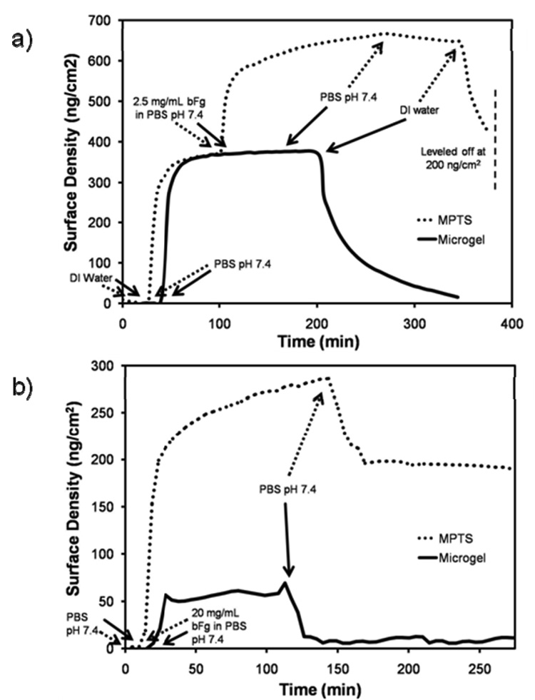
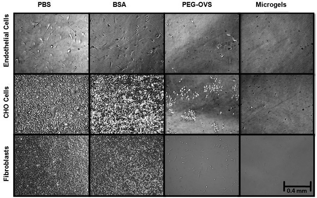
Late-term thrombosis on drug-eluting stents is an emerging problem that might be addressed using extremely thin, biologically active hydrogel coatings. We report a dip-coating strategy to covalently link poly(ethylene glycol) (PEG) to substrates, producing coatings with approximately <100 nm thickness. Gelation of PEG-octavinylsulfone with amines in either bovine serum albumin (BSA) or PEG-octaamine was monitored by dynamic light scattering (DLS), revealing the presence of microgels before macrogelation. NMR also revealed extremely high end-group conversions prior to macrogelation, consistent with the formation of highly crosslinked microgels and deviation from Flory-Stockmayer theory. Before macrogelation, the reacting solutions were diluted and incubated with nucleophile-functionalized surfaces. Using optical waveguide lightmode spectroscopy (OWLS) and quartz crystal microbalance with dissipation (QCM-D), we identified a highly hydrated, protein-resistant layer with a thickness of approximately 75 nm. Atomic force microscopy in buffered water revealed the presence of coalesced spheres of various sizes but with diameters less than about 100 nm. Microgel-coated glass or poly(ethylene terephthalate) exhibited reduced protein adsorption and cell adhesion. Cellular interactions with the surface could be controlled by using different proteins to cap unreacted vinylsulfone groups within the coating.
Figures


Similar articles
-
Ultralow protein adsorbing coatings from clickable PEG nanogel solutions: benefits of attachment under salt-induced phase separation conditions and comparison with PEG/albumin nanogel coatings.Langmuir. 2013 Mar 26;29(12):4128-39. doi: 10.1021/la3051115. Epub 2013 Mar 11. Langmuir. 2013. PMID: 23441808 Free PMC article.
-
Chondroitin sulfate coatings display low platelet but high endothelial cell adhesive properties favorable for vascular implants.Biomacromolecules. 2014 Jul 14;15(7):2512-20. doi: 10.1021/bm5003762. Epub 2014 Jun 25. Biomacromolecules. 2014. PMID: 24927450
-
High salt stability and protein resistance of poly(L-lysine)-g-poly(ethylene glycol) copolymers covalently immobilized via aldehyde plasma polymer interlayers on inorganic and polymeric substrates.Langmuir. 2006 Jun 20;22(13):5760-9. doi: 10.1021/la0602766. Langmuir. 2006. PMID: 16768506
-
Adsorption of proteins at physiological concentrations on pegylated surfaces and the compatibilizing role of adsorbed albumin with respect to other proteins according to optical waveguide lightmode spectroscopy (OWLS).J Biomater Sci Polym Ed. 2013;24(13):1499-518. doi: 10.1080/09205063.2013.772045. Epub 2013 Mar 6. J Biomater Sci Polym Ed. 2013. PMID: 23848445
-
Size-selective protein adsorption to polystyrene surfaces by self-assembled grafted poly(ethylene glycols) with varied chain lengths.Langmuir. 2005 Sep 13;21(19):8774-84. doi: 10.1021/la051049r. Langmuir. 2005. PMID: 16142960
Cited by
-
Enhancing single molecule imaging in optofluidics and microfluidics.Int J Mol Sci. 2011;12(8):5135-56. doi: 10.3390/ijms12085135. Epub 2011 Aug 12. Int J Mol Sci. 2011. PMID: 21954349 Free PMC article. Review.
-
Combining an optical resonance biosensor with enzyme activity kinetics to understand protein adsorption and denaturation.Biomaterials. 2015 Jan;38:86-96. doi: 10.1016/j.biomaterials.2014.10.002. Epub 2014 Nov 1. Biomaterials. 2015. PMID: 25453976 Free PMC article.
-
In vivo tests of a novel wound dressing based on biomaterials with tissue adhesion controlled through external stimuli.J Mater Sci Mater Med. 2011 May;22(5):1357-64. doi: 10.1007/s10856-011-4299-2. Epub 2011 Mar 25. J Mater Sci Mater Med. 2011. PMID: 21437637
-
Interaction of human plasma proteins with thin gelatin-based hydrogel films: a QCM-D and ToF-SIMS study.Biomacromolecules. 2014 Jul 14;15(7):2398-406. doi: 10.1021/bm500750v. Epub 2014 Jul 2. Biomacromolecules. 2014. PMID: 24956040 Free PMC article.
-
Inhibiting Bacterial Adhesion by Mechanically Modulated Microgel Coatings.Biomacromolecules. 2019 Jan 14;20(1):243-253. doi: 10.1021/acs.biomac.8b01378. Epub 2018 Dec 19. Biomacromolecules. 2019. PMID: 30512925 Free PMC article.
References
-
- Jeon S, Lee J, Andrade J, DeGennes P. Protein Surface Interactions in the Presence of Polyethylene Oxide .1. Simplified Theory. Journal of Colloid and Interface Science. 1991;142:149–158.
-
- Elbert D, Hubbell J. Surface treatments of polymers for biocompatibility. Annual Review of Materials Science. 1996;26:365–394.
-
- Kim J, Wacker BK, Elbert DL. Thin polymer layers formed using multiarm poly(ethylene glycol) vinylsulfone by a covalent layer-by-layer method. Biomacromolecules. 2007;8:3682–3686. - PubMed
-
- Lopez GP, Ratner BD, Tidwell CD, Haycox CL, Rapoza RJ, Horbett TA. Glow discharge plasma deposition of tetraethylene glycol dimethyl ether for fouling-resistant biomaterial surfaces. J Biomed Mater Res. 1992;26:415–439. - PubMed
-
- Cao L, Sivaprasad S, Ratner BD, Horbett TA. Glow discharge plasma treatment of polyethylene tubing with tetraglyme results in ultralow fibrinogen adsorption and greatly reduced platelet adhesion. Journal of Biomedical Materials Research Part A. 2006;79A:788–803. - PubMed
Publication types
MeSH terms
Substances
Grants and funding
LinkOut - more resources
Full Text Sources
Other Literature Sources
Miscellaneous
